Purpose
To evaluate sensitivity and specificity of contrast-enhanced ultrasound (CEUS) findings of morphologically confirmed hyperplastic and neoplastic parathyroid glands in patients with hyperparathyroidism.
Methods and materials
Prospective study included 75 patients (18-77 years, F:M=64:11, with hyperparathyroidism scheduled for parathyroidectomy. B-mode ultrasound, Colour Doppler, Superb Microvascular Imaging(SMI), CEUS (SonoVue 1 ml vs. 2ml) images obtained and postprocessing performed. Results compared with postoperative morphology - 68 parathyroid adenomas (PA), 17 parathyroid hyperplasia (PH).It may be challenging to differentiate parathyroid lesions from lymph nodes and thyroid nodules in B-mode US because of visual similarities between these structures, especially in patients with goitres and after previous neck surgery [1-3]. Contrast-enhanced US with intravenous administration of...
Results
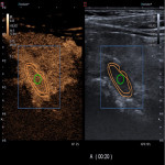
Morphology results indicated 60 solitary parathyroid adenomas (PA), 5 atypically localized lesions. Most characteristic ultrasound features of PA vs. PH were: hypoechoic, well defined lesions with central increased B mode echogenicity (44% and 50%, respectively), peripheral to central vascularisation (47% and 66%, respectively) with feeding vessel (92% for both), average size of adenoma 1456mm3 vs. hyperplasia 899mm3 (p=0.001). CEUS showed peripheral hypervascularity in early arterial phase (median=11s), quickly reaching peak contrast concentration (median=16s), following early washout (median=29s) in PA and homogenous contrasting dynamics in PH....
Conclusion
CEUS of parathyroid lesions shows high sensitivity and specificity in differentiation of adenoma, regardless after injecting 1 ml vs 2 ml of contrast agent. Hypereplasia shows homogeneous enhancement, uptake and washout appears fastpaced in comparson to adenoma, whereas adenoma is characterized by peripheral uptake, central washout and slower hemodynamics.
Personal information and conflict of interest
S. Pavlovics:
Nothing to disclose
M. Radzina:
Nothing to disclose
Z. Narbuts:
Nothing to disclose
M. Miķelsone:
Nothing to disclose
M. Ratniece:
Nothing to disclose
R. Niciporuka:
Nothing to disclose
M. Liepa:
Nothing to disclose
P. Prieditis:
Nothing to disclose
A. Ozolins:
Nothing to disclose
References
1. Uller, W.; Jung, E.; Hornung, M.; Ross, C.; Jung, W.; Schlitt, H.; Stroszczynski, C.; Agha, A. Evaluation of the microvascularization of pathologic parathyroid glands in patients with primary hyperparathyroidism using conventional ultrasound and contrastenhanced ultrasound. Clin. Hemorheol. Microcirc. 2011, 48, 95–103.
2. Sung, J.Y. Parathyroid ultrasonography: The evolving role of the radiologist. Ultrasonography 2015, 34, 268–274.
3. Polat, A.V.; Ozturk, M.; Akyuz, B.; Celenk, C.; Kefeli, M.; Polat, C. The diagnostic value of shear wave elastography for parathyroid lesions and comparison with cervical lymph...


