Keywords:
Head and neck, Ultrasound, Contrast agent-intravenous, Localisation, Endocrine disorders, Neoplasia
Authors:
S. Pavlovics, M. Radzina, Z. Narbuts, M. Miķelsone, M. Ratniece, R. Niciporuka, M. Liepa, P. Prieditis, A. Ozolins
DOI:
10.26044/ecr2022/C-10658
Results
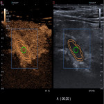
Morphology results indicated 60 solitary parathyroid adenomas (PA), 5 atypically localized lesions. Most characteristic ultrasound features of PA vs. PH were: hypoechoic, well defined lesions with central increased B mode echogenicity (44% and 50%, respectively), peripheral to central vascularisation (47% and 66%, respectively) with feeding vessel (92% for both), average size of adenoma 1456mm3 vs. hyperplasia 899mm3 (p=0.001). CEUS showed peripheral hypervascularity in early arterial phase (median=11s), quickly reaching peak contrast concentration (median=16s), following early washout (median=29s) in PA and homogenous contrasting dynamics in PH. We observed PH homogeneous uptake and washout fastpaced in comparson to PA (p=0.001). The most common histological subtype of adenoma was chief-cell adenoma (87%, n=59). PH showed higher wash-in and wash-out rates both in central and peripheral parts, compared to PA (p=0.001) and higher ACSI values in central parts (p=0.002) and periphery (p=0.001). Higher ACSI was observed in the periprhery among parathyroid adenomas. CEUS had sensitivity and specificity for differentiation of PA of 92% and 69% respectively, PPV 90%, NPV 70% (p=0.05), regardless quantum of contrast media (p=0.1). Sensitivity was 98%, specificity 80%, PPV 98%, NPV 80%, and accuracy was 96.4% after quantitative lesion analysis.


