Keywords:
Artificial Intelligence, CT-High Resolution, Computer Applications-Detection, diagnosis, Tissue characterisation
Authors:
C. H. Leow, V. Corona, P. Yousefi, M. Purtorab, K. Tait, S. Mohammadi, B. Irving, G. Brüggenwerth
DOI:
10.26044/ecr2022/C-14712
Results
Results
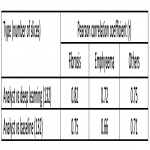
Table 1 illustrates the correlation between the Analyst, Baseline, and our DL approach in fibrosis and emphysema quantification. Quantification using DL approach shows higher correlation with the ground truth compared to baseline approach.
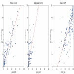
Figure 2 shows the correlation plots for DL vs. ground truth (132 slices). Close clustering around the regression line suggests agreement between DL and ground truth.
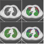
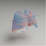
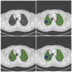
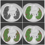
Figure 3-6 show the comparison of each approach to the raw image and ground truth, highlighting the qualitative performance of DL compared to baseline.
A subset of 18 cases was also annotated by the clinical expert, before the analysts' annotations were reviewed. The set was too limited to draw any conclusions from, however the results for interobserver variation are described here. Analyst vs Expert (Fibrosis = 0.72, Emphysema =0.987, Others = 0.69), Expert vs Deep learning (Fibrosis = 0.51, Emphysema =0.68, Others = 0.5).
The results show a strong correlation between ground truth and our DL model for pulmonary fibrosis and emphysema with Pearson's correlation coefficient of 0.82 and 0.72, respectively (as shown in Table 1). Other studies [9] showed a correlation coefficient between observers and their model of an average of 0.37 for the combined IIP pathologies, and 0.24 for emphysema. Our approach shows promise, with higher correlations between ground truth and predicted segmentations. The importance of IIP and fibrosis/emphysema quantification in prediction of clinical outcomes has been shown [11,12,15,16]. The proposed approach could potentially be used to help clinicians in quantifying and monitoring pulmonary fibrosis and emphysema for IIP patients more reliably in clinical studies, as well as in routine settings.


