Type:
Educational Exhibit
Keywords:
Cardiovascular system, Ultrasound-Colour Doppler, Contrast agent-other, Arteriosclerosis
Authors:
A. Lioznovs, M. Radzina, L. Saule, I. Briede
DOI:
10.26044/ecr2022/C-19894
Findings and procedure details
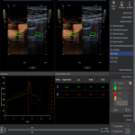
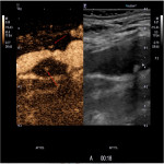
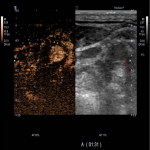
CEUS examination conducted after B mode and Color doppler and Spectral doppler multiparametric ultrasound evaluation by the ultrasound equipment with specific contrast software using contrast agent SonoVue (Bracco, Milan) 1,0-4,8 ml with intravenous administration of contrast bolus followed by saline flush (10 ml). CEUS scanning usually is performed continuously for first 90 seconds and continued intermittently for up to 3-5 minutes. The dynamic process of scanning is stored as cine loops and still images with information about timing. Stored clips are used for later review or specified postprocessing as the examination is dynamic and subtle uptake requires additional analysis.
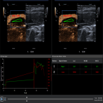
Postprocessing of contrast uptake analysed qualitative, semiquantitative and quantitative (using specified software provided by contrast media manufacturer – VueBox (Bracco, Milan), providing time intensity curves after placement of measurement ROI on the plaque.
Plaque neovascularization can be graded in different categories, one of them is Nakamura et al's7 modified criteria of neovascularization in 3 categories:
- Grade 0 - neovascularization not detected.
- Grade I - neovascularization detected on the side or base of the plaque (spot type or linear).
- Grade II - diffuse neovascularization.
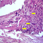
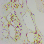
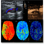
The results of the studies where unstable plaque was detected in 73 patients showed that CEUS and SMI has greater accuracy in the determination of atherosclerotic haemodynamically significant stenosis than in conventional Duplex Doppler ultrasonography. Comparing CEUS method and results of histology statistically significant correlation was found (rs = 0.61; p = 0.002). Statistically significant correlation was found between CEUS and SMI methods (rs = 0,801; p = 0,0001). Comparing results with CEUS grading and contrast arrival time, was found statistically significant difference between groups (p=0.035) with earlier vasa vasorum enhancement in Grade 2 subgroup.
Studies have shown that CEUS is able to provide the functional state of the stent better than classic duplex dopplerography, as well as accurately determine the degree of restenosis and the need for re-treatment for the patient.5,6
Extensive calcinosis is important limitation factor in carotid atherosclerotic plaque neovascularization diagnostics by CEUS, reducing methods sensitivity by 24.45%, specificity 22.5%, negative predictive value up to 2 times, positive predictive value by 16.24%.

