Learning objectives
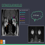
Review of the orbital anatomy and cranial nerves involved in eye movement.
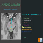
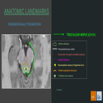
Critical anatomic landmarks of each neural pathway
Radiologic assessment: optimal MRI protocol.
Etiologic assortment according to topographic division.
Key radiological findings of representative cases within our hospital setting.
Background
Diplopia means double vision and is the simultaneous perception of two images when observing a single object with both eyes open. These images can have a horizontal, vertical or oblique disposition.There are 2 types of diplopia:
Monocular diplopia: secondary to ocular globe pathologies.
Binocular diplopia: secondary to ocular misalignment due to impaired extraocular muscle function (ophtalmoplegia or ophtalmoparesia). It is caused by lesions located inside the orbital cavity or along the course of oculomotor (CN III), trochlear (CN IV) and abducens (CN VI) cranial nerves....
Findings and procedure details
IMAGING ASSESMENT OF BINOCULAR DIPLOPIA
MRI is the recommendend imaging modality in the evaluation of patients with binocular diplopia because of its high image resolution in the assesment of the brainstem, subarachnoid spaces, cavernous sinus and orbital structures. It is critical to perform an MRI study in the following cases: new onset diplopia in patient less than 50 years old, presence of more than one neurologic symptom, symptom progression or history of cancer.
Optimal MRI protocol should include:
axial T2w of brain and brainstem
axial/coronal...
Conclusion
Binocular diplopia can be caused by potentially life-threatening entities hence the vital importance of recognizing the anatomy of the neural courses of CN III, IV and VI.A thorough understanding of the patient setting as well as the spectrum of clinical presentations of ocular dysmotility is needed to provide a careful neuroimaging assessment so the quality of patient care can significantly improve when clinical and radiological findings correlate.
Personal information and conflict of interest
A. Micolich Vergara:
Nothing to disclose
B. Beltrán Mármol:
Nothing to disclose
A. Gené Orriols:
Nothing to disclose
M. Saint-Gerons:
Nothing to disclose
J. M. Maiques Llácer:
Nothing to disclose
References
Kim, J. H., Kim, M., & Bae, Y. J. (2022). Magnetic Resonance Imaging in Diplopia: Neural Pathway, Imaging, and Clinical Correlation. Korean journal of radiology, 23(6), 649–663. https://doi.org/10.3348/kjr.2022.0101
Kirsch, C. F., & Black, K. (2017). Diplopia: What to Double Check in Radiographic Imaging of Double Vision. Radiologic clinics of North America, 55(1), 69–81. https://doi.org/10.1016/j.rcl.2016.08.008
Thatcher, J., Chang, Y. M., Chapman, M. N., Hovis, K., Fujita, A., Sobel, R., & Sakai, O. (2016). Clinical-Radiologic Correlation of Extraocular Eye Movement Disorders: Seeing beneath the Surface. Radiographics :...


![Fig 4: Subacute left paramedian midbrain stroke → left CN III palsy
62 year-old female with a history of alcohol abuse and bipolar disorder, presents with left eye ptosis, intermittent diplopia with partial limitation to elevate and adduct left eye together with right hemiparesis and ataxia. A brain MRI and contrast enhanced MR angiography was performed showing a focal lesion in the left paramedian midbrain which is hyperintense on T2w FLAIR and DWI [a,b d], with normal ADC values [c], which suggests a subacute stroke. MR angiography did not show any abnormalities in anterior or posterior circulation.
Origin: Department of Radiology, Hospital del Mar – Parc de Salut Mar, Barcelona, Spain/2022](https://epos.myesr.org/posterimage/esr/ecr2023/164532/media/940244?maxheight=150&maxwidth=150)
![Fig 5: Chronic left midbrain hemorrhagic lesion affecting CN III nucleus.
80 year-old male, with a history of hypertension treated with four different anti-hypertensive drugs, type 2 diabetes and atrial fibrillation. Presents with dysarthria, gait ataxia with right lateralisation and vertical diplopia with inability to adduct and elevate the left eye.
Initial brain CT scan shows small hyperdense lesion located at left parasagittal midbrain [a] and brain MRI depicts the same lesion which is isointense on T1w sequence [c], low signal intensity on T2w GE and Venous BOLD sequences [d,e] and has hyperintense surrounding edema on T2 FLAIR [b].
As a result of this lesion the patient presented with developed decreased vision together with bilateral optic nerve atrophy, therefore underwent a 2 year follow-up brain and orbit MRI which showed fatty infiltration of extraocular muscles affecting predominantly the superior, lateral and inferior rectus muscles on T1w [g,h], STIR [i] and T1w FLAIR sequences [f] and increase in perineural space surrounding both optic nerves suggesting mild atrophy.
Origin: Department of Radiology, Hospital del Mar – Parc de Salut Mar, Barcelona, Spain](https://epos.myesr.org/posterimage/esr/ecr2023/164532/media/934618?maxheight=150&maxwidth=150)
![Fig 6: Subacute hemorrhagic pineal lesion.
36 year-old male with no previous history of morbidity or trauma, presents with binocular blurry vision, headache and horizontal diplopia triggered with left gaze. Brain MRI with gadolinium demonstrated a pineal hemorrhagic lesion represented by hyperintensity on T1w [b,c], low signal intensity on T2 GE with presence of hematocrit level [a] and mild peripheral gadolinium-enhancement on T1w C+(Gd) [d]. After 3 months, follow up brain MRI depicted resolution of hemorrhagic components showing a cystic pineal lesion with high signal on T2w [g] and thin peripheral enhancement on T1w C+ [e] composed of fluid-fluid level and a hypointense anterior structure (probably calcification) on T2w GE [f].
Origin: Department of Radiology, Hospital del Mar – Parc de Salut Mar, Barcelona, Spain](https://epos.myesr.org/posterimage/esr/ecr2023/164532/media/934619?maxheight=150&maxwidth=150)
![Fig 7: Stage IV Burkitt-like lymphoma invading cavernous sinus → left CN VI paralysis
72 year-old male with multiple cardiovascular risk factors presents with onset of binocular diplopia.
Brain MRI revealed the presence of a solid mass occupying the left cavernous sinus and part of Meckel’s cave that is isointense to soft tissue on T1w and T2w sequences [a,b]. This mass surrounds the carotid siphon on T2w FLAIR sequences [c] without causing stenosis and has a slight gadolinium-enhancement on T1w C+ sequences [d]
Origin: Department of Radiology, Hospital del Mar – Parc de Salut Mar, Barcelona, Spain](https://epos.myesr.org/posterimage/esr/ecr2023/164532/media/934620?maxheight=150&maxwidth=150)
![Fig 8: Orbital fracture and post-operative frozen eye → left CN VI paralysis
28 year-old male that suffers a motorbike accident. Admission brain CT showed a large left periorbital hematoma with displaced left orbital floor fracture, anterior maxillary sinus wall fracture and displaced nasal bone fracture with associated hemosinus [a,b]. On coronal view we can see displaced bone fragments with orbital fat and inferior rectus muscle prolapse into maxillary sinus without signs of entrapment [c,d].
The patient underwent orbital floor surgical repair [e,f] and developed onset of left eye abduction restriction. Follow-up CT depicted thickening of left lateral rectus muscle without signs of entrapment [g].
Origin: Department of Radiology, Hospital del Mar – Parc de Salut Mar, Barcelona, Spain](https://epos.myesr.org/posterimage/esr/ecr2023/164532/media/934621?maxheight=150&maxwidth=150)
![Fig 9: Left orbital cavernous malformation.
54 year-old female without any history of morbidity, presentes with left eye proptosis and diplopia. Brain MRI with Gd revealed a large, rounded, well-circumscribed left orbital mass that molds into the intraconal space causing moderate proptosis and superolateral displacement of the optic nerve without infiltrating it [b,c,d ]. This mass is isointense to the muscles in T1w [a ], hyperintense on T2w [e] and presents a slow and gradual heterogeneous post-gadolinium enhancement [f,g ].
Origin: Department of Radiology, Hospital del Mar – Parc de Salut Mar, Barcelona, Spain/2022](https://epos.myesr.org/posterimage/esr/ecr2023/164532/media/940245?maxheight=150&maxwidth=150)
![Fig 10: Graves orbithopathy decompensation → thickening of left inferior rectus muscle
64 year-old male with a history of smoking and Graves disease, presents with progression of associated orbitopathy with binocular vertical diplopia and strabismus decompensation.
Brain and orbit MRI demonstrated bilateral exoftalmos [a ] with mild thickening of left inferior rectus muscle [d] alongside fatty infiltration of all EOM predominantly affecting both inferior rectus muscles, as shown on T1w [c ], T1w FS [d] and STIR sequences [e].
Origin: Department of Radiology, Hospital del Mar – Parc de Salut Mar, Barcelona, Spain/2021](https://epos.myesr.org/posterimage/esr/ecr2023/164532/media/940246?maxheight=150&maxwidth=150)
![Fig 11: Craniofacial fibrous dysplasia (left orbit posterolateral wall (zygomatic bone and sphenoid wing) → left RL muscle
59 year-old male with a history of smoking and obesity, presents with chronic frontal headache and horizontal diplopia with inability to abduct the left eye.
Brain and orbit MRI was performed revealing the presence of a bony expansion of the diploe affecting the posterolateral wall of the left orbit (zygomatic bone and sphenoid wing) on 3D T1w FSE [a,d] causing mild obliteration of intraconal space [b,c] and slight medial displacement of left RL muscle [a].
Origin: Department of Radiology, Hospital del Mar – Parc de Salut Mar, Barcelona, Spain/2020](https://epos.myesr.org/posterimage/esr/ecr2023/164532/media/940247?maxheight=150&maxwidth=150)
