ESSR 2016 / P-0019
Radiology trainee reporting of out of hours cervical spine trauma CT scans:do they make mistakes? Does it matter?
Congress:
ESSR 2016
Poster Number:
P-0019
Type:
Scientific Poster
Keywords:
Musculoskeletal spine, CT, Computer Applications-General, Acute
Authors:
S. B. Gagrani1, R. Bhatt2, S. Lee3, N. Khan3; 1Birmingham/UK, 2Leicester /UK, 3Leicester/UK
DOI:
10.1594/essr2016/P-0019

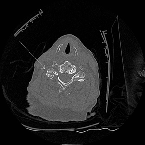
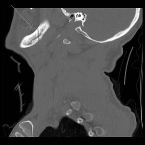
Fig. 1:
Axial CT image of fracture through the right C4/5 facet joint with widening.

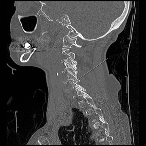
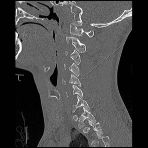
Fig. 2:
Sagittal CT image of fracture through the right C4/5 facet joint.

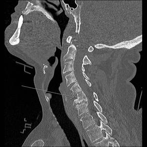
Fig. 3:
Sagittal CT image of fracture through the left C5 foramen transversarium and...

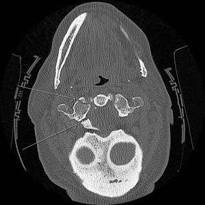
Fig. 4:
Axial CT slice demonstrating a fracture through the right C5 foramen...

Fig. 5:
Coronal CT image showing an undisplaced fracture through the left occipital...

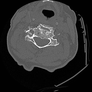
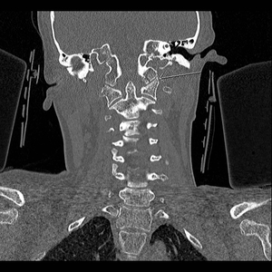
Fig. 6:
Axial CT slice of an acute fracture of the right lateral mass of C1 vertebra.

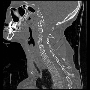
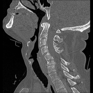
Fig. 7:
Sagittal CT slice demonstrating widening of the interspinous distance between...

Fig. 8:
Sagittal image of 40% anterior wedge compression of C6 vertebral body.

Fig. 9:
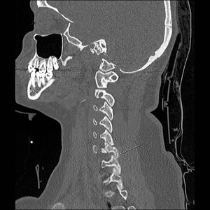
Sagittal CT Slice demonstrating partially united fracture mandible at the...

Fig. 10:
Sagittal CT slice of undisplaced fracture of the right transverse process of C5

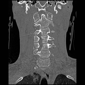
Fig. 11:
Coronal CT of undisplaced fracture of the inferior articular process of C3 on...

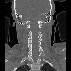
Fig. 12:
Coronal CT showing abnormal widening of the left C6/C7 facet joint with...

Fig. 13:
Sagittal CT image demonstrates abnormal widening of the left C6/C7 facet joint...

Fig. 14:
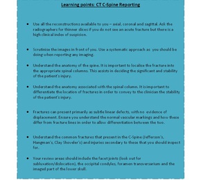
How to assess a CT of the C-Spine