
ESSR 2017 / P-0184
Glomangiomas of skeleton and soft tissues demystified
Congress:
ESSR 2017
Poster Number:
P-0184
Type:
Educational Poster
Keywords:
Authors:
R. Vadapalli1, H. Vardhan1, S. Jha1, A. S. Vadapalli2; 1Hyderabad/IN, 2Pune AFMC/IN
DOI:
10.1594/essr2017/P-0184

Fig. 1:
Dynamic contrast enhanced T1 weighted MRI distal phalanx of little finger...

Fig. 2:
MR Imaging features of Glomus tumours

Fig. 3:
Subungual glomus tumour radiography:Saucerization and marginal erosion seen ...

Fig. 4:
CE MRA images showing the intensely enhancing Glomangioma in nail bed of index...

Fig. 5:
Time resolved CE MRA technique showing delayed phase images of enhancing blush...

Fig. 6:
Young 17 year old female with intense pain in third toe with tenderness on...

Fig. 7:
CE MRA images showing the intensely enhancing Glomangioma in nail bed of third...

Fig. 8:
CE MRA images showing the intensely enhancing Glomangioma in nail bed of third...

Fig. 9:
CE MRA images showing the intensely enhancing Glomangioma in nail bed of third...

Fig. 10:
Intra op images showing the exposed nail bed in subungual portion 3rd toe

Fig. 11:
Vascular glomus in toe nail bed subungual zone -Exposed and resected with...

Fig. 12:
Multiple glomangiomas : Dynamic post contrast enhancing big toe planar surface...

Fig. 13:
Young boy with severe pain and distress with tenderness on touch in infra...

Fig. 14:
Essentially normal looking except for a subtle soft tissue thickening anterior...

Fig. 15:
Cone down soft tissue radiography with Low KV technique reveals well defined...
 shows a mixed echogenicity mass in infra patellar location
References: rammohan vadapalli Hyderabad India")
Fig. 16:
Ultrasound imaging:Ultrasound with doppler(colour images not shown) shows a...

Fig. 17:
Intra op images at the infra patellar location exposing vascular soft tissue...

Fig. 18:
Intra op images at the infra patellar location exposing vascular soft tissue...

Fig. 19:
Intra op images at the infra patellar location with resected vascular soft...

Fig. 20:
Glomangioma infra patellar subcutaneous soft tissues

Fig. 21:
Glomangioma infra-patellar subcutaneous soft tissues: Cut section of specimen...

Fig. 22:
Glomangioma infra patellar subcutaneous soft tissues Histopathology
 in distal phalanx
References: rammohan vadapalli Hyderabad India")
Fig. 23:
Plain radiographs in subungual radiographs show osteopenia secondary to...

Fig. 24:
Glomangioma of thumb –subungual zone of distal phalanx –Intra operative...
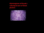
Fig. 25:
Glomangioma infra patellar subcutaneous soft tissues
Blood vessels surrounded...

Fig. 26:
Glomangio-sarcoma of thumb:Proximal phalanx of thumb expansile lytic intra...

Fig. 27:
Glomangio-sarcoma of thumb:CE MRA with Volume rendering showing neo...

Fig. 28:
Glomangio-sarcoma of thumb:
Features of glomangiosarcomas may include the...