ESSR 2018 / P-0066
A step-by-step imaging tour of congenital abnormalities of the foot
Congress:
ESSR 2018
Poster Number:
P-0066
Type:
Educational Poster
Keywords:
Congenital, Normal variants, Plain radiographic studies, MR, CT, Musculoskeletal system
Authors:
B. Sharif, M. Khoo; Stanmore/UK
DOI:
10.1594/essr2018/P-0066

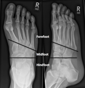
Fig. 1:
Right foot radiographs demonstrating the broad anatomical divisions of the foot...

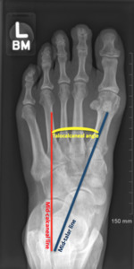
Fig. 2:
Weight bearing AP radiograph of the left foot demonstrating normal hindfoot...

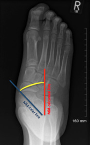
Fig. 4:
Weight bearing radiograph of the right foot. There is hindfoot valgus, the...

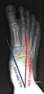
Fig. 3:
Weight bearing AP radiograph of the right foot demonstrating a reduced...

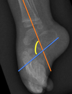
Fig. 5:
An alternative way of measuring the talocalcaneal angle using a lateral...

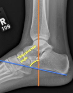
Fig. 6:
Lateral radiograph of the right ankle demonstrating how to measure the...
 due to the foot being held in fixed equinus.")
Fig. 7:
Lateral radiograph of the right foot demonstrating an increased tibiocalcaneal...