This study includes every MRI performed in the pediatric population (0-21 years old) between May 2016 and May 2018 at our institution,
for evaluation of musculoskeletal conditions.
In total,
1000 pediatric MRIs were performed for the purpose of evaluating musculoskeletal disease in 891 patients.
Circa 54% (N=540) of patients were female and 46% (N=460) male.
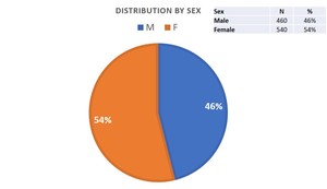
Fig. 1: Pediatric MRI examinations - distribution by sex
The most examined segments were knee (35,1%),
hip (17%),
ankle (8,8%),
toes and feet (5,3%),
hands and fingers (4,5%),
shoulder (4,3%) and whole-body/muscle (3,1%).

Table 1: Pediatric MRI examinations - distribution by body segment
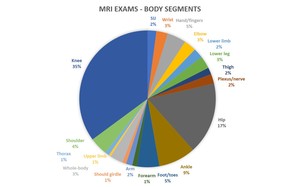
Fig. 2: Pediatric MRI examinations - distribution by body segment
Average age was 11,62 years,
which was higher for evaluation of sacro-iliac (14,33 yo),
knee (13,52 yo),
ankle (12,36 yo) and wrist (12,26 yo) joints.
Average was was lowest on plexus/nerve examinations (5,04 yo),
forearm (8,01 yo),
arm (8,85 yo) and lower limb (8,86 yo) segments.
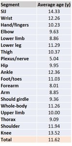
Table 2: Pediatric MRI examinations - average age by body segment
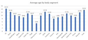
Fig. 3: Pediatric MRI examinations - average age by body segment
Regarding frequency of main diagnoses,
distribution was as follows:
- Inflammation and/or infection – 18,2%
- Normal examination – 16%
- Soft tissue trauma - 13,8%
- Tumor – 11%
- Undetermined/nonspecific – 8,3%
- Bony trauma – 6,6%
- Musculoskeletal malformation - 4,8%
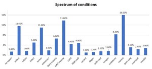
Fig. 4: Pediatric MRI examinations - spectrum of conditions
Whole-body (and shoulder girdle) MRIs had the highest percentage of normal examinations (38,7% and 44,4%,
respectively),
followed by thoracic (40%) segments,
sacro-iliac (42,8%) and hip (19,4%) joints.
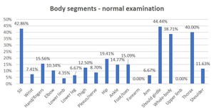
Fig. 5: Pediatric MRI examinations - normal examinations by body segment
Regarding nonspecific or undetermined findings,
whole-body,
forearm,
upper limb and thoracic examinations had the highest frequency (see table below).
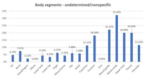
Fig. 6: Pediatric MRI examinations - undetermined/nonspecific examinations by body segment
Signs of inflammation as the main imaging finding was common (11,6% of all examinations) and most frequent on sacro-iliac joint (38,1%),
elbow (20,7%),
arm and upper limb (20%) examinations.
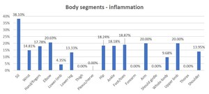
Fig. 7: Pediatric MRI examinations - inflammation by body segment
Interestingly,
the sacro-iliac joints had two main diagnoses - either normal examination (42,9%) or inflammation (38,1%).
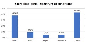
Fig. 8: Pediatric MRI examinations - Sacro-iliac joint spectrum of conditions
Other body segments and their spectrum of conditions are depicted below.
A few highlights include:
- bony trauma of the wrist was the most common finding,
followed by inflammation and soft tissue cyst
- musculoskeletal malformations were described in 20% of hand and finger examinations,
followed by inflammatory and normal findings (17,8% and 15,6%,
respectively)
- bony trauma of the elbow was the most common finding (38%),
as well as inflammation and infection (20,69%)
- inflammatory changes were reported as the main finding in 18,2% of hips; 19,4% of hip examinations were normal
- soft tissue trauma and inflammatory findings of the ankle were the most common diagnoses (19,3% and 18,2% ,
respectively),
followed by normal examinations in a significant proportion (14,8%)
- shoulder examinations showed a significant proportion of bony and soft tissue trauma (18,6% and 16,3%,
respectively)
- By far the most common diagnosis in the knee was soft tissue trauma (28,2%),
followed by normal examinations (14,8%)
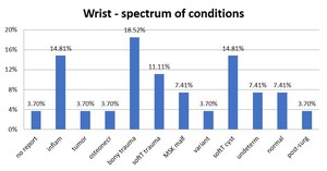
Fig. 9: Pediatric MRI examinations - Wrist joint spectrum of conditions
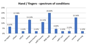
Fig. 10: Pediatric MRI examinations - Hand/fingers spectrum of conditions
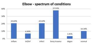
Fig. 11: Pediatric MRI examinations - Elbow joint spectrum of conditions
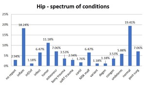
Fig. 12: Pediatric MRI examinations - Hip joint spectrum of conditions
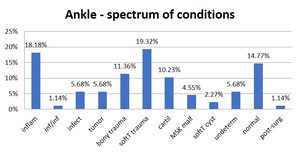
Fig. 13: Pediatric MRI examinations - Ankle joint spectrum of conditions
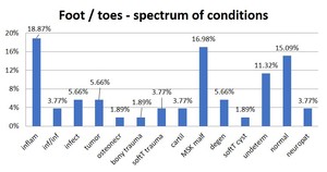
Fig. 14: Pediatric MRI examinations - Foot/toes spectrum of conditions
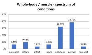
Fig. 15: Pediatric MRI examinations - Whole-body/muscle spectrum of conditions
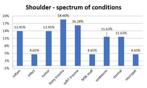
Fig. 16: Pediatric MRI examinations - Shoulder joint spectrum of conditions
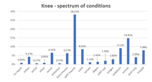
Fig. 17: Pediatric MRI examinations - Knee joint spectrum of conditions
Focus on specific groups of conditions was made possible given the large amount of data collected.
Of note, 38% of tumors found were benign soft tissue tumors and 44% corresponded to benign bone tumors.
Circa 14% of bone tumours presented with aggressive features.
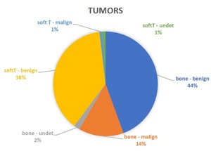
Fig. 18: Pediatric MRI examinations - Soft tissue and bone tumors
Limitations and future perspectives
- It was not possible to extract clinical motive for examination as the information was not available in a significant proportion of patients
- In some cases,
more than one condition was present,
and the most relevant for the purpose of the study was selected (example: bony trauma and soft tissue trauma commonly co-exist,
and bony trauma was given more relevance due to its clinical implications)
- Creation of a database/atlas for normal MRIs of different musculoskeletal segments and for different age groups
- Creation of a database for most common,
uncommon and some rare findings and conditions
- Evaluation of MRI protocols,
image quality and problems faced,
to employ targeted methodologies to improve diagnosis and efficiency
