Objective measures of image quality are available [8,
9,
13].
Quantitative CT image quality metrics allow for standardized and reproducible quantitative image quality analysis,
e.g.
for routine quality assurance (QA),
inspection of image quality discrepancies between different scanner models and acquisition protocols,
and evaluation of new CT technology or reconstruction methods,
cf.
Fig. 4.

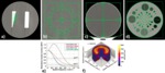
Fig. 4: Determination of CT image quality (IQ) metrics such as modulation transfer function (MTF), noise-power spectrum (NPS), noise magnitude, CT number accuracy, uniformity across the field-of-view (FOV), contrast-to-noise ratio (CNR), and signal-to-noise ratio (SNR) of simulated lesions in phantoms allows for standardized and reproducible quantitative IQ analysis, e.g. for routine quality assurance (QA), inspection of IQ discrepancies between different scanner models and acquisition protocols, and evaluation of new CT technology or reconstruction methods. Examples of cross-sectional CT image slices of phantoms used for quantitative IQ determination and exemplary results: (a) MTF, (b) three-dimensional (3D) NPS, (c) noise magnitude, CT number accuracy, and uniformity across the field-of-view (FOV), (d) contrast-to-noise ratio (CNR) and signal-to-noise ratio (SNR) of simulated lesions, (e) MTFs and (f) 3D NPS determined using phantom image data as shown in (a) and (b), respectively [13]. Note that despite being fully quantitative, these image quality metrics do not enable direct judgement of whether the achieved IQ is appropriate for enabling reliable and reproducible diagnosis with regard to each specific diagnostic question encountered in clinical routine.
References: Pahn et al. (2016) Toward standardized quantitative image quality (IQ) assessment in computed tomography (CT):�A comprehensive framework for automated and comparative IQ analysis based on ICRU Report 87. Phys Med 32 (1) [13].
However,
despite being fully quantitative,
experience shows that such objective measures of image quality cannot lay claim to a complete description of images in view of their diagnostic quality,
i.e.
with regard to all of the features relevant to making a correct clinical diagnosis [5,
6].
Therefore,
quantitative image quality metrics do not enable direct judgement of whether the achieved image quality is appropriate for enabling reliable and reproducible diagnosis with regard to each specific diagnostic question encountered in clinical routine [13].
The subjective assessment of image quality by radiologists can very easily differ from an objectively determined order of ranking [5].
Determining the image quality “appropriate” for a certain diagnostic purpose can be a complex task,
as both quantitative metrics (e.g.
noise,
spatial resolution,
etc.) and observer perceptions (fine structure discernibility and low-contrast detail detectability) are involved [6],
cf.
Fig. 5.

Fig. 5: Illustration of image quality (IQ) dependence on reconstruction method and radiation exposure for the example of high-resolution CT (HR-CT) imaging of the lungs: (a – c) have been acquired at standard dose (tube potential 120 kVp, tube current-time product 180 mAs, gantry rotation time 0.5 s, collimation 128 × 0.6 mm, helical pitch 0.993, effective dose Deff = 8.18 mSv); (d – f) have been acquired at low dose with low tube potential and current-time product (80 kVp, 60 mAs, other settings as for (a – c)), resulting in one tenth of radiation exposure (Deff = 0.85 mSv). Applying different reconstruction methods on the same CT raw data yields distinctly different IQ: (a) & (d) Filtered backprojection (FBP) exhibits highest noise, (b) & (e) statistical iterative reconstruction (iDose4, Philips Healthcare, Best, The Netherlands) reduces noise and suppresses artifacts, (c) & (f) iterative model-based reconstruction (IMR, Philips Healthcare, Best, The Netherlands) is virtually noise- and artifact-free, but seemingly leads to a wash-out of fine lung details at ultra-low dose. Experimental data of porcine lungs in mid-inspiratory breath hold have been acquired in the same animal (anaesthetized and mechanically ventilated) in an in-vivo experiment.
Nonetheless,
it should be possible to identify what “appropriate image quality” for commonly performed CT examinations is,
and to define “appropriate image quality” in terms of spatial resolution,
low-contrast detectability and image noise on a “per-organ” basis in view of diagnostic question.
To date,
published data from digital radiography indicates that is indeed possible to define standardized quality criteria,
e.g.
based on common orthopedic assessments necessary for therapeutic decisions and therapy monitoring,
that might not only be useful for overall radiation exposure reduction but as general criteria for “appropriate image quality” [10].
Definitions of diagnostic task-based “appropriate image quality” should be driven by the clinicians’ requirements of everyday clinical routine and needs input from the radiologists’ community by subspecialty.
Ideally,
the task-based “appropriate image quality” criteria derived and driven by clinical practice can subsequently be used for retrospective automatic analysis of acquired image data on a “per-exam” basis directly after completion of each imaging examination in view of whether “appropriate image quality” is achieved,
exceeded,
or not satisfactorily met.
To this end,
model observers that objectively assesses image quality for clinically relevant tasks,
e.g.
as described in ICRU Report No.
54,
need to be developed [7].
As,
for example,
human observers' performance in view of expected low contrast detectability can be predicted by model observers,
these represent a promising method in terms of radiologists' sensitivity performance and are therefore of relevance in the clinical environment [14,
15].

 dependence on reconstruction method and radiation exposure for the example of high-resolution CT (HR-CT) imaging of the lungs: (a – c) have been acquired at standard dose (tube potential 120 kVp, tube current-time product 180 mAs, gantry rotation time 0.5 s, collimation 128 × 0.6 mm, helical pitch 0.993, effective dose Deff = 8.18 mSv); (d – f) have been acquired at low dose with low tube potential and current-time product (80 kVp, 60 mAs, other settings as for (a – c)), resulting in one tenth of radiation exposure (Deff = 0.85 mSv). Applying different reconstruction methods on the same CT raw data yields distinctly different IQ: (a) & (d) Filtered backprojection (FBP) exhibits highest noise, (b) & (e) statistical iterative reconstruction (iDose4, Philips Healthcare, Best, The Netherlands) reduces noise and suppresses artifacts, (c) & (f) iterative model-based reconstruction (IMR, Philips Healthcare, Best, The Netherlands) is virtually noise- and artifact-free, but seemingly leads to a wash-out of fine lung details at ultra-low dose. Experimental data of porcine lungs in mid-inspiratory breath hold have been acquired in the same animal (anaesthetized and mechanically ventilated) in an in-vivo experiment.")