Materials:
- Siemens Somatom Sensation 64 CT scanner
- Alderson Rando anthropomorphic phantom
- Tissue equivalent slabs of resin
- Radcal 10x06-6 ion chamber (Calibrated Sept 2019, c = 0.99)
Methodology:
The RANDO phantom produced by Alderson Research Laboratories together with slabs of tissue equivalent resin were used to simulate a pregnant patient. The RANDO phantom is separated in to thirty five sections: sections 1-34 are 2.5 cm thick and section 35 is 9 cm thick.
A calibrated Radcal 10x6-6 ion chamber was placed in the following positions 6 for dose measurement:
Trimester 1 = 6 cm from pubic symphysis at a depth of 6.3 cm
Trimester 2 = 20.5 cm from pubic symphysis at a depth of 7.3 cm
Trimester 3 = 34.5 cm from pubic symphysis at a depth of 8.3 cm
The set up for trimester 3 is shown in figure 1.
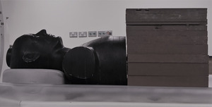
Fig. 1: Trimester 3 set-up
Figure 1: Trimester 3 set-up.
The simulated pregnant patient was scanned with the standard Radiology Dept. CT protocols presented in Table 1.
|
Protocol
|
Trimester
|
kV
|
mAs
|
FOV
|
DLP mGy.cm
|
CTDIvol mGy
|
|
CTPA
|
1
|
100
|
118
|
M
|
156.12
|
5.26
|
|
2
|
100
|
128
|
M
|
169.33
|
5.70
|
|
3
|
100
|
170
|
M
|
292.64
|
7.59
|
|
CT Abdo
|
1
|
120
|
102
|
L
|
407.03
|
7.87
|
|
2
|
120
|
92
|
L
|
226.02
|
7.11
|
|
3
|
120
|
183
|
L
|
631.07
|
14.02
|
|
Brain
|
3
|
120
|
360
|
S
|
427.99
|
47.55
|
Table 1: Radiology Dept CT examination protocols
Results:
Fetal dose.
|
Protocol
|
Trimester
|
Dose (mGy)
|
Error (mGy)
|
|
Brain
|
1
|
0
|
±0.01
|
|
2
|
0
|
±0.01
|
|
3
|
0
|
±0.01
|
|
CTPA
|
1
|
0.07
|
±0.01
|
|
2
|
0.15
|
±0.01
|
|
3
|
3.9
|
±0.08
|
|
Abdomen-Pelvis
|
1
|
4.2
|
±0.08
|
|
2
|
7.5
|
±0.01
|
|
3
|
8.3
|
±0.02
|
Table 2. Estimates of radiation dose to the fetus for three common CT examinations for each trimester.
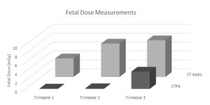
Fig. 2: Results
Discussion:
A CT brain scan of the simulated pregnant patient produced no measurable response from the ionisation chamber (threshold 100 nGy) for any of the three trimesters of pregnancy. It can therefore be concluded that the dose to the fetus from a brain scan during any period of gestation is negligible. This result supports the published literature on this issue2.
The CTPA protocol produced a fetal dose estimate of 0.07±0.01 mGy for trimester one, 0.15±0.01 mGy for trimester two and 3.90±0.08 mGy for the third trimester simulation. Finally CT of the abdomen of a pregnant patient with the fetus in the field of view produced doses of 4.18±0.08 mGy, 7.53±0.01 mGy and 8.27±0.02 mGy for trimesters one to three respectively.
Fetal doses measured in this study are significantly less than the 100 mGy minimum threshold for the occurrence of radiogenic malformations in the fetus and at the measured doses, the probability of inducing cancer is low7.
Limitations:
The study needs to be repeated on other CT scanners and the measured doses verified using other radiation dose measurement methods.
