Normal Urinary Bladder
- The fetal urinary bladder is well visualized during the 1st trimester.
In many instance,
however,
the bladder is not visualized during the US exam without definite abnormality
- Although the fetus normally fills and empties bladder every 30 to 45 minutes, urinary bladder is visualized during the US exam in the 2nd and 3rd trimester
- The bladder wall is very thin and located in the central pelvis.
Bilateral internal iliac arteries run around the bladder and branch the umbilical arteries,
which is the differential finding of normal bladder from other intra-abdominal cystic lesions

Fig. 17
References: 1Department of Radiology, Seoul National University Hospital

Fig. 18
References: Department of Radiology, Seoul National University Hospital/Republic of Korea 2012
Large Urinary Bladder : 1st Trimester
- The normal size of the urinary bladder During the 1st trimester is smaller than 7mm
- Megacystis during the 1st trimester usually associated with bladder outlet obstruction such as posterior urethral valve and cloacal malformation
- Posterior urethral valve occurs in the male only
- Almost cloacal malformation occurs in female
- The outcome of bladder outlet obstruction is usually poor without proper treatment,
and is associated with other fetal anomalies in many cases
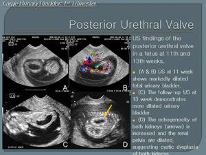
Fig. 2
References: Department of Radiology, Seoul National University Hospital/Republic of Korea 2012

Fig. 1
References: Department of Radiology, Seoul National University Hospital/Republic of Korea 2012
Invisible Urinary Bladder: 2nd & 3rd Trimester
- Invisible bladder during the 2nd & 3rd trimester usually needs careful radiological evaluation of the fetus and estimation of amniotic fluid volume
- Invisible bladder with severely decreased amniotic fluid means impairment of urine production caused by bilateral renal agenesis,
bilateral MCDK,
autosomal recessive polycystic kidney,
and syndromes such as sirenomelia and Meckel-Gruber syndrome
- Invisible bladder with normal amniotic fluid may be associated with bladder exstrophy
1> Invisible urinary bladder with Anhydramnios

Fig. 3
References: Department of Radiology, Seoul National University Hospital/Republic of Korea 2012

Fig. 4
References: Department of Radiology, Seoul National University Hospital/Republic of Korea 2012

Fig. 5
References: Department of Radiology, Seoul National University Hospital/Republic of Korea 2012

Fig. 6
References: Department of Radiology, Seoul National University Hospital/Republic of Korea 2012

Fig. 7
References: Department of Radiology, Seoul National University Hospital/Republic of Korea 2012
2> Invisible urinary bladder with Normal Amniotic fluid Volume
Invisible fetal urinary bladder with normal amniotic fluid is common condition due to fetal urination.
In most cases the bladder is filled within 30 minutes. The bladder exstrophy is the result of a deficiency in development of the lower abdominal wall musculature,
so that the bladder is open and the mucosa of the bladder is continuous with the skin.
Bladder cannot contain the urine and is invisible on the prenatal image.
However kidneys are usually normal and the volume of amniotic fluid is normal

Fig. 8
References: Department of Radiology, Seoul National University Hospital/Republic of Korea 2012
Large Urinary Bladder: 2nd & 3rd Trimester

Fig. 9
References: Department of Radiology, Seoul National University Hospital/Republic of Korea 2012

Fig. 10
References: Department of Radiology, Seoul National University Hospital/Republic of Korea 2012
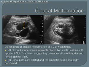
Fig. 11
References: Department of Radiology, Seoul National University Hospital/Republic of Korea 2012
Other Abnormal findings of Urinary bladder
1> Ureterocele

Fig. 12
References: Department of Radiology, Seoul National University Hospital/Republic of Korea 2012
2> Cyst around Urinary bladder
Many cystic lesions around the fetal urinary bladder,
including ovarian cyst,
duplication cyst,
seminal vesicle cyst,
MCDK of pelvic kidney can develop during the fetal life

Fig. 13
References: Department of Radiology, Seoul National University Hospital/Republic of Korea 2012
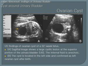
Fig. 14
References: Department of Radiology, Seoul National University Hospital/Republic of Korea 2012
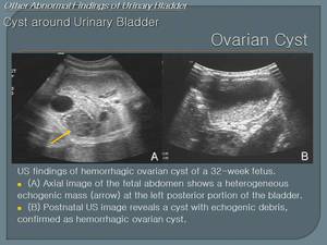
Fig. 15
References: Department of Radiology, Seoul National University Hospital/Republic of Korea 2012
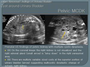
Fig. 16
References: Department of Radiology, Seoul National University Hospital/Republic of Korea 2012


