RESULTS:
CT scan*Unilateral polypoid mass
-Hypodense (n=20)
-Heterogeneous enhancement (n=20)
-Associated with bone erosion (n=12)
*Location:
-Nasal wall (n=15)
-Maxillary sinus (n=4)
-thmoid complex (n=1)
Coronal CT scan showed a soft-tissue mass involving the right nasal fossa (cross), with contiguous extension into maxillary sinus and ethmoidal cells . Ethmoidal infundibulum is enlarged.
Note sclerosis of lateral and inferior walls of maxillary sinus, and thinning of the inferior wall of the orbit. The tumor erode the lamina papyracea
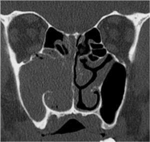
Fig.: Coronal CT scan showed a soft-tissue mass involving the right nasal fossa, with contiguous extension into maxillary sinus and ethmoidal cells . Ethmoidal infundibulum is enlarged. Note sclerosis of lateral and inferior walls of maxillary sinus and thinning of the inferior wall of the orbit.
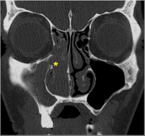
Fig.: Coronal CT scan showed a soft-tissue mass involving the right nasal fossa (cross), with contiguous extension into maxillary sinus and ethmoidal cells . Ethmoidal infundibulum is enlarged. The tumor erode the lamina papyracea
Axial and coronal CT scan showed a large right nasal mass beneath middle meatus, with extension through eroded medial wall of the maxillary sinus, and scalloping of his postero lateral wall.
Note erosion of the nasal septum and extension to the left nasal fossa.
Extension through the choana into the nasopharynx
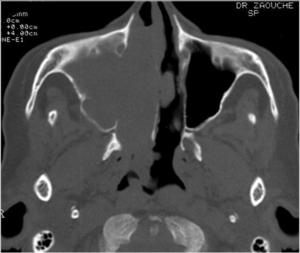
Fig.: Axial CT scan showed a large right nasal mass beneath middle meatus with extension through eroded medial wall of the maxillary sinus, and scalloping of his postero lateral wall.Extension through the choana into the nasopharynx
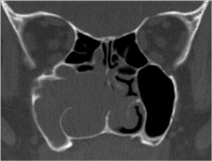
Fig.: Coronal CT scan showed a large right nasal mass beneath middle meatus, with extension through eroded medial wall of the maxillary sinus.Note erosion of the nasal septum and extension to the left nasal fossa
Axial and coronal CT scan showed a focal lobulated tumor, involving the left spheno-ethmoidal recessus
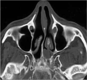
Fig.: Axial CT scan showed a focal lobulated tumor, involving the left spheno-ethmoidal recessus
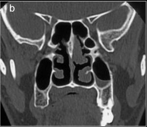
Fig.: Coronal CT scan showed a focal lobulated tumor, involving the left spheno-ethmoidal recessus
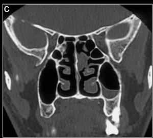
Fig.: Coronal CT scan showed a focal lobulated tumor, involving the left spheno-ethmoidal recessus
MRI
*Signal abnormalities:
-Intermediate on T1 and T2 n=10
-Hypo T1, Hyper T2 n=4
-Hyper T1, Hypo T2 n=2
*Lobulated aspect n=8
*Solid heterogenous enhancement n=16
Axial T2 and T1 post-gadolinium weighted MRI images showing a large mass involving the nasal fossa and the right maxillary sinus, with a columnar aspect.
Note the difference between tumor(hyposignal T2 and heterogeneous enhancement) and retained secretions (hypersignal T2 and peripheral
enhancement)
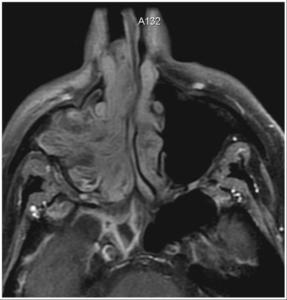
Fig.: Axial T2 weighted MRI images showing a large mass involving the nasal fossa and the right maxillary sinus, with a columnar aspect
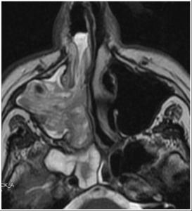
Fig.: Axial T1 post-gadolinium weighted MRI images showing a large mass involving the nasal fossa and the right maxillary sinus, with a columnar aspect Note the difference between tumor (hyposignal T2 and heterogeneous enhancement) and retained secretions (hypersignal T2 and peripheral enhancement)
*Staging system by Krouse*
-T1: n=1
-T2: n=6
-T3: n=7
-T4: n=2
Coronal, sagittal T2 and axial T1 post-gadolinium weighted MRI showed a soft-tissue mass involving the right maxillary sinus and the nasal fossa, presenting a columnar aspect, and with aheterogeneous enhancement
Sagittal plane allow the radiologist to estimate extension to nasopharynx
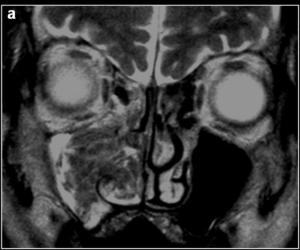
Fig.: Coronal T2 weighted MRI showed a soft-tissue mass involving the right maxillary sinus and the nasal fossa, presenting a columnar aspect
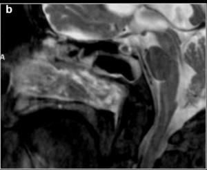
Fig.: Sagittal T2 weighted MRI showed a soft-tissue mass involving the right maxillary sinus and the nasal fossa, presenting a columnar aspectSagittal plane allow the radiologist to estimate extension to nasopharynx
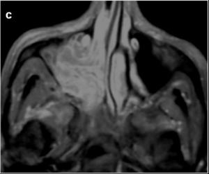
Fig.: axial T1 post-gadolinium weighted MRI showed a soft-tissue mass involving the right maxillary sinus and the nasal fossa presenting a heterogeneous enhancement
Management-Endoscopic approach in all patients
-Adjunctive open approach performed in 2 patients (Mini Caldwell-Luc)
-Incomplete excision in 3 patients
Follow-up: ranged from 4 months to 6 years and half (mean: 3 years)
Recurrence: 3 patients
-4 months after surgery: n=1
-5 months after surgery: n=1
-30 months after surgery: n=1
Association to carcinoma: n=1
Errors in MRI staging-Overestimation of extension to frontal sinus in 3 patients (T3vsT2)
-Inflammation of extra orbital fat without tumor involvement (T4vs T2)
-Squamous cell carcinoma diagnosed at histologic examination (T3vs T4)
Inverted papilloma of the right nasal fossa with typic aspect on MRI
(T3 by Krouse). Malignant cells find on histology
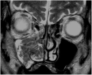
Fig.: Inverted papilloma of the right nasal fossa with typic aspect on MRI (T3 by Krouse). Malignant cells find on histology
CASE REPORTS
CASE 1: A 74-year old man presented with chronic nasal obstruction and rhinorrhea. Endoscopic examination showed a left nasal tumor extended to nasopharynx
Axial CT scan showed a soft-tissue mass involving the left nasal fossa and ethmoid cells (cross) with retained secretions in maxillary and sphenoid sinuses. Note the presence of calcifications(arrow)
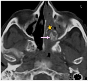
Fig.: Axial CT scan showed a soft-tissue mass involving the left nasal fossa and ethmoid cells (cross) with retained secretions in maxillary and sphenoid sinuses.Note the presence of calcifications (arrow)
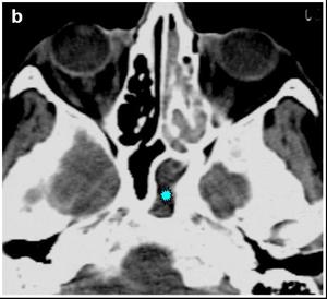
Fig.: Axial CT scan showed a soft-tissue mass involving the left nasal fossa and ethmoid cells with retained secretions in maxillary and sphenoid sinuses (cross)
Coronal CT images of the same patient
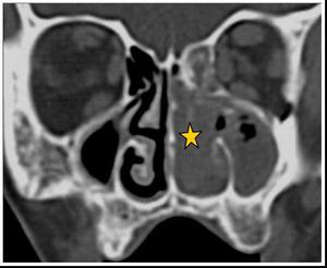
Fig.: Coronal CT image showed the tumour (cross)
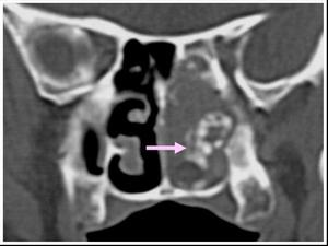
Fig.: Coronal CT images showed calcifications (arrow)
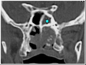
Fig.: Coronal CT image showed retained secretions in sphenoid sinus (cross)
He had an endoscopic approach
3 years after, recurrence of rhinorrhea with epistaxis
Coronal CT images showed tumor recurrence involving left spheno-ethmoidal recessus
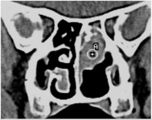
Fig.: Coronal CT image showed tumor recurrence involving left spheno-ethmoidal recessus
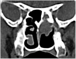
Fig.: Coronal CT image showed tumor recurrence involving left spheno-ethmoidal recessus
He had open approach
Absence of recurrence 3 years after
Case 2: a 40-year old man presented with left nasal occlusion and rhinorrhea.
On clinical examination a diagnosis of inverted papilloma was made.
Axial CT scan showed a mass involving the left nasal fossa, with extension to ethmoid cells and maxillary sinus.
Note the erosion of middle turbinate
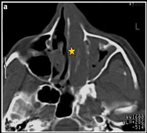
Fig.: Axial CT scan showed a mass involving the left nasal fossa, with extension to ethmoid cells and maxillary sinus (cross).Note the erosion of middle turbinate
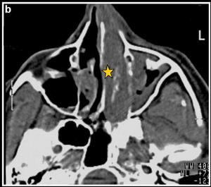
Fig.: Axial CT scan showed a mass involving the left nasal fossa, with extension to ethmoid cells and maxillary sinus (cross).
Coronal CT images showed the extension to the orbit through the lamina papyracea.
Note the erosion of the nasal septum
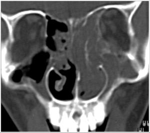
Fig.: Coronal CT image showed the extension to the orbit through the lamina papyraceaNote the erosion of the nasal septum
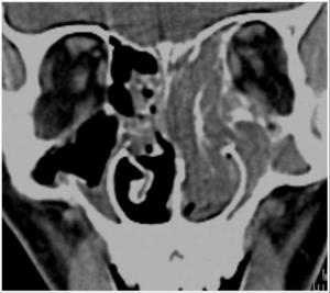
Fig.: Coronal CT image showed the extension to the orbit through the lamina papyracea Note the erosion of the nasal septum (open arrow)
The patient had an endoscopic approach
2 years after, recurrence of rhinorrhea and anosmia
Axial and coronal CT scan showed a mass in the original tumor location
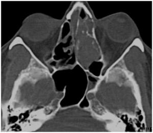
Fig.: Axial CT scan showed a mass in the original tumor location
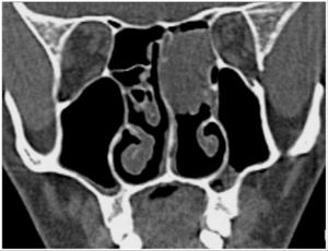
Fig.: Coronal CT scan showed a mass in the original tumor location
He had an open approach (latero-nasal and frontal)
4 years after, second recurrence.
Coronal, axial T2 and axial post-gadolinium T1 with fat sat showed a recurrence of inverted papilloma in the ethmoid complex (cross), presenting an important and heterogeneous enhancement
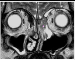
Fig.: Coronal T2 showed a recurrence of inverted papilloma in the ethmoid complex
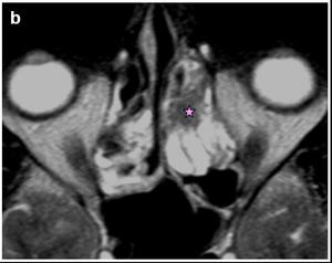
Fig.: Axial T2 showed a recurrence of inverted papilloma in the ethmoid complex
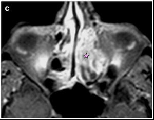
Fig.: axial post-gadolinium T1 with fat sat showed a recurrence of inverted papilloma in the ethmoid complex, presenting an important and heterogeneous enhancement (cross)
The patient had a second endoscopic approach, with no recurrence after 12 months
DISCUSSION:
Epidemiology:-Rare benign neoplasm: 0,5-4%
-Reported in all age groups from adolescence to older life, peak incidence in the fifth and sixth decades
-In older patients: associated carcinoma+++
-Male to female ratio of 3:1 ( 10:1 in our serie)
Etiology:-Unknown
-Association with human papillomavirus HPV 11
HPV 16 associated with carcinoma
-Traumatic, allergic and environmental etiologies suspected
Pathology:-Inversion of the neoplastic epithelium into the underlying stroma
-Basement membrane is respected
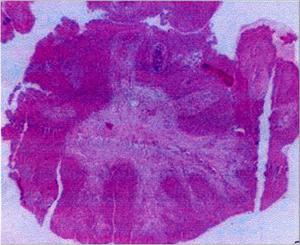
Fig.: Inversion of the neoplastic epithelium into the underlying stroma
-Unilateral nasal obstruction: most common presenting symptom (66-100%)
-Rhinorrhea: 22-66%
-Epistaxis: 22-52% : predictive symptom of malignancy if associated with pain
-Symptom duration: 2 weeks -20 years
Endoscopic examination:-Firm, lobulated, translucid, friable and often hemorrhages when manipulated tumor
-Arising from the lateral nasal wall usually in the region of the middle meatus (96%)
-Involving the paranasal sinuses: ethmoid complex+++, maxillary sinus++
CT scan:-Distinctive but not pathognomonic
-Polypoid, hypodense mass (66%*), filling the nasal fossa, presenting a -heterogeneous enhancement
-Calcifications: 52% of cases
*Entrapped bone
*Tumoral calcifications
-Modifications of adjacent bone:
*Thinning
*Erosion (7-50%)
*Bowing
*Sclerosis: hyperplastic reaction to the chronic sinusitis
-Inflammatory reaction is present in 90%* of cases: homolateral in 86%, controlateral in 40%
-Limits:
*Distinguishing between inflammation and neoplasm
*Non accurate tumor delineation
MRI:-No signature pattern of MRI signal intensity characteristics or enhancement
-Columnar aspect present in 50% of our patients
-Lobulated contours
-Heterogeneous enhancement
-Distinguishing between:
*Tumor: Hypo T2, heterogeneous and important enhancement
*Inflammation: Hyper T2, peripheric enhancement
-Accurate tumor delineation: extension to frontal and sphenoidal sinuses, to orbit and cerebrum
-Coincidence between MRI and postoperative staging:
*Oikawa: 86%
*Our study: 68,7%
-MRI overestimated extension in frontal sinus, orbit and cerebrum in our study
RECURRENCE:-After open approach: 8*-17%**
-After endoscopic approach: 10*-14%**
-Our study: 13%
-Explications:
*Incomplete excision+++
*Multicentric origin
-Location:
*Site of original tumor: maxillary and ethmoid sinuses
*Sphenoid sinus, cribriform plate and frontal sinus were involved at proportionately higher rates in comparison to their incidence of initial invovement
-MRI: first imaging modality in cases of patent recurrence or doubtful endoscopy
-Similar appearences to primary tumor
-Lobulated aspect++
-Low to intermediate signal on T1 and T2 weighting with heterogeneous enhancement
-CT: limited in distinguishing between postoperative inflammation and recurrence
-90% of recurrences occur within 5 years*
-No correlation between number of recurrences and development of malignancy
MALIGNANCY:-5-15%
-Association to HPV 16
-Old patients > 65 years
-Epistaxis, pain...
-Synchronous or metachronous
-Multiple recurrences++
-Histology: squamous cell carcinoma+++
-MRI: non specific:
*Intense and homogeneous enhancement
*Important bone erosion
OTHER DIAGNOSIS:-Antrochoanal polyps: teenagers and young adults, enlargement of infundibulum
-Aspergillosis: Hypo T1 and T2 not modified after Gadolinium, Calcifications++
-Mucoceles, juvenile nasopharyngeal angiofibroma...
-Squamous cell carcinoma
