Embryology of the diaphragm
The diaphragm is formed through the fusion of tissue from four different sources
- The septum transversum,
a thick mass of mesoderm between the primitive heart tube and the developing liver,
gives rise to most of the central tendon
- The paired pleuroperitoneal membranes are sheets of somatic mesoderm that develop from the dorsal and dorsolateral body wall
- The dorsal mesentery of the oesophagus is invaded by myoblasts and forms the crura of the diaphragm
- The body wall contributes muscle to the peripheral portions of the definitive diaphragm
Figure 1

Fig.: Embryology of the diaphragm
References: Restrepo CS, Eraso A, Ocazionez D, Lemon J, Martinez S, Lemons DF. The diaphragmatic crura and retrocrural space: Normal imaging appearance, variants, and pathologic conditions. RadioGraphics 2008; 28:1289-1305.
- ST – septum transversum
- ppm – pleuroperitoneal membranes
- dme – dorsal mesentery of the oesophagus
- Bw – body wall
- IVC – inferior vena cava
- Es – oesophagus
- Ao - Aorta
Anatomy of the diaphragm and crura
The diaphragm is a dome-shaped fibromuscular septum which separates the thoracic and abdominal cavities
Its peripheral part consists of muscular fibres which take origin from the circumference of the thoracic outlet and converge to be inserted into a central tendon
The muscular fibres may be grouped according to their origins into three parts; sternal,
costal,
and lumbar
- The sternal part arises by two fleshy slips from the back of the xiphoid process
- The costal part from the inner surfaces of the cartilages and adjacent portions of the lower six ribs on either side,
interdigitating with the transversus abdominis
- The lumbar part from aponeurotic arches,
named the lumbocostal arches,
and from the lumbar vertebrae by the two diaphragmatic crura
Figure 2
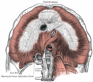
Fig.: Diaphragm from below
References: Gray, Henry. Anatomy of the Human Body. Philadelphia: Edinburgh, Scotland: Churchill Livingstone, 2000.
The diaphragmatic crura
The crura are strong tendons attached to the anterolateral surfaces of the upper lumbar vertebrae and blend with the anterior longitudinal ligament of the vertebral column
- The right crus, larger and longer than the left,
arises from the anterior surfaces of the bodies of the upper three lumbar vertebrae
- The left crus arises from the corresponding parts of the upper two lumbar vertebrae only
Muscle fibres radiate from each crus,
diverge and pass superiorly before curving anteriorly into the central tendon
Tendinous fibres from the medial edge of each crus unite,
anterior to the aorta,
at the level of T12 to from the median arcuate ligament
Figure 3

Fig.: Axial contrast enhanced CT demonstrating the diaphragmatic crura and retrocrural space
The retrocrural space
The retrocrural space is bounded by:
- Anteriorly – the median arcuate ligament
- Anterolaterally – the right and left crus
- Posteriorly – vertebral bodies
Figure 4
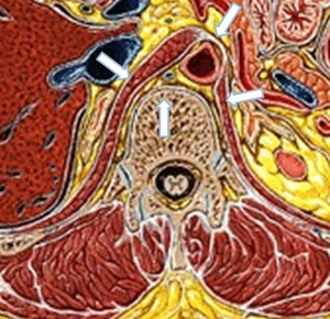
Fig.: Boundaries of the retrocrural space
References: www.netterimages.com
Normal contents of the retrocrural space
The normal retrocrural space contains fatty tissue,
the aorta,
nerves,
veins of the azygos system,
lymph nodes,
cisterna chyli and the thoracic duct
Figure 5

Fig.: Axial contrast enhanced CT outlining the normal anatomy of the retrocrural space and its contents
Aorta
The aorta is the largest structure within the retrocrural space
At the level of the aortic hiatus,
the aorta is slightly left of midline
Within the retrocrural space,
the aorta gives off posterior intercostal and subcostal arterial branches
Figures 6 and 7

Fig.: Axial and coronal contrast enhanced CT’s showing the normal aorta and its position within the retrocrural space
Azygos and hemiazygos veins
Azygos vein
- The azygos vein is usually formed by the union of the ascending lumbar and subcostal veins of the right side
- It passes through the aortic opening under or through the right crus
Hemiazygos vein
- The hemiazygos vein is formed by the joining of the left ascending lumbar and subcostal veins
- It passes under cover of or through the left crus
