This poster was previously presented in Spanish at the 2010 Congreso Nacional SERAM (A Coruña)
Keywords:
Head and neck, Eyes, Trauma, CT, MR, Ultrasound, Diagnostic procedure, Foreign bodies, Neoplasia
Authors:
M. A. Martin Perez1, J. M. Millán Juncos2, R. Blanco Hernández1, I. Martín García1, C. Martinez Lara1; 1Zamora/ES, 2Madrid/ES
DOI:
10.1594/ecr2012/C-0715
Methods and Materials
We describe the radiological manifestations using CT and MR of orbital traumatic injuries,
suspected or foreign body, tumor pathology of the eye (melanoma, choroidal osteoma) and intraconic space primary disease (lymphoma, meningioma, optic nerve glioma,
hemangioma), metastatic or secondary infiltration vicinity of facial and intracranial invasive lesions.
Radiological findings are illustrated in vascular and inflammatory disease of the extraocular muscles, including basic processes such as myositis, Brown syndrome,
or thyroid ophthalmopathy. Also in this section we emphasize the contribution of imaging in the evaluation of the different causes of exophthalmos.
In our work,
we conducted a systematic classification of the pathology,
broken down by location:
- Facial bones and orbital walls (Continent).
- Orbital content
- Surrounding structures (who suffer disease that secondarily affect the orbit)
![](https://epos.myesr.org/posterimage/esr/ecr2012/110428/media/381681?maxheight=300&maxwidth=300)
Fig. 3: 
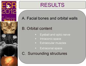
Results. Classification.