Keywords:
Abdomen, Pancreas, MR, CT, Diagnostic procedure, Cysts
Authors:
C. Delavaud1, G. D'ASSIGNIES1, J. Cros1, P. Ruszniewski1, P. Hammel1, A. Couvelard2, V. Vilgrain1, M.-P. Vullierme1; 1Clichy sur Seine/FR, 2Paris/FR
DOI:
10.1594/ecr2013/B-0576
Results
Clinical and biological findings
Clinical and biological findings of patients with ACC and BD-IPMN are described in table 1 :
|
Patient
|
ACC n=5
|
BD-IPMN n=20
|
p value
|
|
Clinical features
|
|
|
|
|
Sex (Female)
|
4 (80%)
|
12 (60%)
|
0,4
|
|
Age (years)
|
38,8 (8.01)
|
57,9 (13,30)
|
0.0059
|
|
Personal past history
|
1 Rheumatoid arthritis
|
1 duodenal adenocarcinoma
|
-
|
|
1 breast cancer
|
|
Familial history
|
1 Pancreatic adenocarcinoma
1 pulmonar carcinoma
1 diabetes
|
1 pancreatic
adenocarcinoma
|
-
|
|
Diabetes
|
1 (20%)
|
0
|
0,04
|
|
Exocrine pancreatic failure
|
0
|
0
|
-
|
|
Symptoms
|
|
|
|
|
Pancreatic pain
|
4 (80%)
|
11 (55%)
|
0,30
|
|
Acute pancreatitis
|
2 (40%)
|
11 (55%)
|
0,54
|
|
Incidental finding
|
1 (20%)
|
9 (45%)
|
0,30
|
|
Jaundice
|
0
|
0
|
-
|
|
Others
|
1 polyarthralgia
|
0
|
-
|
|
Follow up (in years)
|
6,25 (4,27)
|
4,9 (1,59)
|
0,26
|
In ACC group,
one patient was lost of follow up and four patients were followed from 1 to 11 years (mean: 6.25 years).
All patients were alive without malignant transformation and without apparition of new cystic lesion after surgery.
Imaging findings
ACC appeared as simple cysts with thin wall on CT and MRI (fig 4).
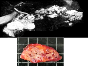
Fig. 4: Radio-pathological correlation of ACC
No tissular thickening or enhancement was found.
Adenopathy or extra-pancreatic invasion was never seen.
In one case,
5 years imaging follow up before surgery was available without any modification.
Imaging findings are summarized in table 2 :
|
|
Patients with ACC n=5
|
Patients with BD-IPMN n=20
|
Odds Ratio (95%CI)
|
P value
|
|
Cyst calcifications
|
4 (80)
|
0
|
48.4 (4.8-∞)
|
0.001
|
|
More than five cysts
|
4(80)
|
4 (20)
|
16 (1.4–185)
|
0.02
|
|
Close peripheral small cysts
|
4 (80)
|
0 (0)
|
48.4 (4.8-∞)
|
0.001
|
|
Size of the biggest cyst
mean (SD)
|
19.8 (13.5)
|
18.3 (9.5)
|
1.01 (0.9– 1.12)
|
0.95
|
|
Cyst in the head (including uncinate process)
|
5 (100)
|
13 (65)
|
3.2 (0.4- ∞)
|
0.27
|
|
Absence of communication with main pancreatic duct
|
3 (75)
|
3 (15)
|
17.0 (1.3– 22,3)
|
0.03
|
|
Absence of main
|
5 (100)
|
14 (70)
|
2.5 (0.3-∞)
|
0.29
|
|
Main pancreatic duct enlargement
|
|
Elongated shape
|
2 (40)
|
12 (60)
|
0.4 (0.06– 3.3)
|
0.42
|
Four variables were statistically associated with ACC in univariate analysis :
- Close peripheral small cysts (fig 5)
- More than five cysts
- Cyst calcifications (fig 6)
- Absence of communication with main pancreatic duct.
Fig. 5: "Close peripheral small cysts pattern" : numerous small cysts present in a small portion of parenchyma and peripheral in location (grey arrow)
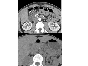
Fig. 6: Cysts calcifications in two cases of ACC
Based on those four variables a score was then computed. Table 3 displays the sensitivity and the specificity of the different cut-off levels of the score :
|
Cutpoint
|
|
|
Sensitivity (%)
|
Specificity (%)
|
|
at least one finding
|
100
|
60
|
|
at least two findings
|
100 |
85
|
|
at least three findings
|
60
|
100 |
|
four findings
|
40
|
100
|

