Introduction
– Large pelvic masses in women may originate from the peritoneum, retroperitoneum, ovaries,
Fallopian tubes, uterus,
cervix,
gastrointestinal tract or bladder.
– To distinguish ovarian from non-ovarian masses is a common imaging dilemma and presents a special diagnostic challenge.
– Diagnosis can be often suggested on the basis of tumour location and anatomic landmarks.
– These landmarks include the organ of origin,
relationship to vasculature,
peritoneal or extraperitoneal involvement and lateral pelvic wall involvement.
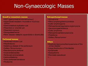
Fig. 1: In this table are enumerate the different non-gynaecological pelvic masses.
References: Department of Radiology, Instituto Português de Oncologia de Lisboa Francisco Gentil, Lisbon/PT
Normal Ovary – Anatomy
– Located in ovarian fossa / fossa of Waldeyer
– Not covered with peritoneum
– Each ovary is suspended in the pelvic peritoneal cavity by three anchoring structures:
- mesovarium – anchors the ovary to the posterior surface of the broad ligament
- utero-ovarian ligament (ovarian ligament) – anchors the ovary to the uterus
- suspensory ligament – anchors the ovary to the pelvic sidewall
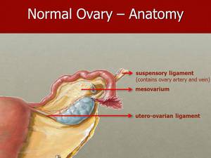
Fig. 2: Illustration of ovarian ligaments.
References: Department of Radiology, Instituto Português de Oncologia de Lisboa Francisco Gentil, Lisbon/PT
– The utero-ovarian ligament and the suspensory ligament of the ovary have variable degrees of laxity.
– They behave more like mesenteries than as tightly fixating or rigid support structures.
– Actual position and orientation of the ovaries are variable (even in the same patient at different times).
– Ovaries may assume unusual locations in the upper pelvis or lower abdomen.
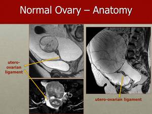
Fig. 3: The presence of ascites allows a clear view of the anchoring structures if the ovaries, such as in these images, where the utero-ovarian ligament can be seen, connecting the ovaries to the uterus.
References: Department of Radiology, Instituto Português de Oncologia de Lisboa Francisco Gentil, Lisbon/PT
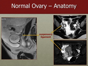
Fig. 4: Suspensory ligaments of the ovaries anchor the ovaries to the pelvic side walls, allowing the passage of the corresponding vessels and nerves.
References: Department of Radiology, Instituto Português de Oncologia de Lisboa Francisco Gentil, Lisbon/PT

