
ECR 2015 / C-0860
Cement pulmonary embolism after balloon kyphoplasty: experience with 68 patients
This poster is published under an open license. Please read the disclaimer for further details.
Congress:
ECR 2015
Poster Number:
C-0860
Type:
Scientific Exhibit
Keywords:
Interventional non-vascular, Musculoskeletal spine, Lung, CT, Digital radiography, Fluoroscopy, Vertebroplasty, Outcomes analysis, Statistics, Outcomes, Osteoporosis, Drugs / Reactions
Authors:
A. A. Fares1, I. Willekens1, S. Alard1, S. A. Z. Khodair2, C. G. Boulet1, M. Moens1, M. De Maeseneer1, J. de Mey1; 1Brussels/BE, 2Qwuesna/EG
DOI:
10.1594/ecr2015/C-0860
,")
Fig. 4:
Distribution of the available postoperative chest radiological investigations...
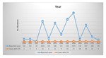
Fig. 5:
Chart-diagram for the most important case series in literature
(All case...