Aims and objectives
Allusions of Х-rays application in anthropology are known from publications since the beginning of the XX-th century.
Nowadays modern visualization techniques are used increasingly for the remains examinations.
However,
information about an assessment of anthropological material using such Х-ray techniques,
as digital microfocus radiography with direct multiple images magnification (DMFR) or multislice computed tomography (MSCT) is limited by a few publications [1–5,
8–11].
With the advent of cone beam computed tomography (CBCT) systems of a new generation it has become possible to conduct researches of...
Methods and materials
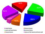
All the anthropological materials were provided by Research Institute and Museum of Anthropology named after D.
N.
Anuchin of Lomonosov Moscow State University (fig.
1).
In total,
24 skeleton fragments,
which were introduced by the soldiers’ remains bone material of the Imperial Napoleon Bonaparte`s army who died in 1812 war,
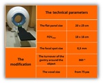
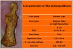
have been examined on modern CBCT-scanner – NewTom 5G (QR S.r.l.,
Italy).
There were a number of indisputable advantages that were applied when cone beam unit had been selected: its modification and technical parameters,
first...
Results
Received CBCT-images of all the anthropological finds were distinguished by high-resolution with a detailed mapping of bone structure: accurate differentiation and direction of bone trabeculae (fig.
5).
It became possible to measure the thickness of the cortical bone,
even if it was less than 1 mm and the length of the defects in those places where it was destroyed.
During the comparative analysis it was found that visualization of bone structure on CBCT-images was comparable or even exceeded MSCT and digital microfocus X-ray images.
In...
Conclusion
The obtained data during the comparative analysis proves the necessity of using modern X-ray techniques with a wide range of the image processing capabilities to detect various types of the bone fractures in anthropological finds.
CBCT-images are comparable with MSCT and it could be recommended as a specific X-ray method of the bone structure posttraumatic intravital and postmortem changes visualization,
which size is even less than 1 – 2 mm.
References
1.
Buzhilova A.
P.,
Berezina N.
Ja.,
Selezneva V.
I. New findings from the collection of Rokhlin: roentgenological analysis of samples from the Paleopatological Fund of MAE RAS.
Electronic library of Peter the Great Museum of Anthropology and Ethnology (the Kunstkamera Museum) of RAS http://www.kunstkamera.ru/lib/rubrikator/08/08_02/978-5-88431-238-8/ (in Russian).
2.
Buzhilova A.
P.,
Dobrovol'skaja M.
V.,
Mednikova M.
B.,
Potrahov N.
N.,
Potrahov E.
N.,
Grjaznov A.
Ju. Application of microfocus roentgenography in diagnostics of diseases of an ancient man.
Peterburgskij zhurnal jelektroniki — St.Petersburg Journal of...


