ECR 2016 / C-1612
Mediastinum, reviewing the epidemiologic aproach: More than lines, bands and interfaces
This poster is published under an open license. Please read the disclaimer for further details.
Congress:
ECR 2016
Poster Number:
C-1612
Type:
Educational Exhibit
Keywords:
Thorax, Mediastinum, Respiratory system, Conventional radiography, CT, MR, Statistics, Epidemiology, Education and training, Neoplasia
Authors:
E. Salinas, L. K. Cifuentes, D. Sossa, B. Pinzon; Bogotá/CO
DOI:
10.1594/ecr2016/C-1612
")
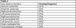
Fig. 1:
Frequency of lines, bands and mediastinal interfaces (3)