A radiation awareness demonstrator system has been implemented in an experimental interventional room at IHU Strasbourg,
containing a robotized X-ray imaging device.
It relies on multiple registered RGBD cameras mounted to the ceiling of the room,
along with radiation simulation and visualization approaches (described below), for providing real-time visual feedback about the current propagation of scattered radiation and the patient and attending personnel dose.
The views from the ceiling cameras are employed to perceive the current environment; this information is then applied by the system for providing consistent visualizations.
Furthermore,
a direct communication with the robotized angiographic C-arm's (Siemens' Artis Zeego) application programming interface (API),
enables the system to have access to the device's current kinematic parameters.
These are applied to display radiation safety information corresponding to the actual C-arm configuration and imaging protocol.
Its graphical user interface (GUI),
depicted in figure 2,
is divided in two main parts.
The left part shows the live feed from the ceiling-mounted cameras,
and the right one shows a virtual representation of the operating room (OR) lay-out.
The imaging device's current kinematic parameters are applied to update the pose of the displayed virtual C-arm model accordingly and also to display radiation safety information corresponding to the actual C-arm configuration.
In a typical usage of the system,
its GUI is shown on a surgical screen inside the operating room for the user to interact with it and be able to choose among the different visualization modes through the icons located on its lower part.
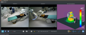
Fig. 2: Graphical User Interface (GUI) of our radiation awareness prototype system.
References: Loy Rodas N., 2018. Context-aware radiation protection for the hybrid operating room. PhD Dissertation, University of Strasbourg, France.
Simulation of X-ray radiation
We have proposed a radiation simulation approach [4,
5] taking the current room lay-out into account to compute in real-time the patient and staff dose,
along with the 3D propagation of scattered radiation.
It applies Monte Carlo methods to compute the propagation of X-rays and the deposited dose for a given imaging protocol and room lay-out.
Our radiation simulation approach exploits the computing capabilities of GPUs to achieve quasi real-time performance in the simulation of the 3D propagation of scattered radiation and patient dose.
In the system’s current version,
the radiation maps are pre-computed for different sets of imaging parameters and angulations,
and are loaded upon the initialization of the system.
They are computed for several standard X-ray particles' energy spectrums (simulated from X-ray tube voltage and filtration values),
for every 5 degree projection in a full C-arm rotation in both LAO/RAO and CAUD/CRAN planes.
Online,
the current C-arm angulation is obtained directly from its API and the corresponding radiation map to display is loaded.
Since for now only kinematic information is provided by the current API's version,
the user selects through the GUI the X-ray imaging parameters to visualize.
Augmented reality visualization of ionizing radiation
Our system can help improving radiation safety by making ionizing radiation visible through different visualization modes [4,
6].
These visualizations rely on our multi-camera setup and tracking/registration methods,
along with the radiation simulation approaches.
We propose to use AR for giving a user the capability to visualize the current patient/staff dose and the 3D distribution and intensity of scattered radiation.
Intraoperatively,
this can contribute to increase radiation awareness and reduce overexposure risks for both patients and staff.
Also,
clinicians can adapt their positioning and the disposition of the protective equipment thanks to the provided visual feedback.
Preoperatively,
these visualizations have the potential to be included into training tools to teach about radiation behavior and about the best safety practices intuitively.
The visualization modes are updated in quasi real-time as the C-arm parameters change to illustrate the effects that altering the projection angle and/or the imaging protocol have on the behavior of radiation.
Two types of color-coded visualizations are provided: a visualization of information shown over a virtual representation of the room (right part of the GUI) and an AR visualization achieved by overlaying registered virtual elements over the color images from the ceiling-mounted cameras (left part).
Visualization in a virtual environment
Relevant information related to the current radiation propagation is shown in a virtual environment.
This enables the system to be also applied in rooms without ceiling-mounted cameras if access to the imaging device's API is available.
The point-of-view of the virtual visualization can be modified by the user for looking at the scene from different perspectives.
As it can be observed in figures 2 and 3,
3D models of the C-arm and operating table,
the 3D propagation of scattered radiation and the 3D position of the persons in the room are displayed.
Furthermore,
the 3D dose deposition over the patient's organs and skin is shown in an additional virtual visualization window.
As depicted in figure 3,
the user can open such a visualization through the system's GUI and the displayed patient exposure map is updated according to current imaging projection and protocol.
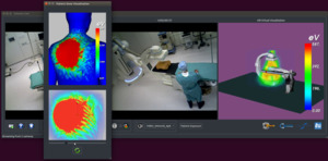
Fig. 3: Visualization of the patient's dose to the internal structures and to the skin for the current X-ray imaging device's projection and imaging protocol on our radiation awareness system.
References: Loy Rodas N., 2018. Context-aware radiation protection for the hybrid operating room. PhD Dissertation, University of Strasbourg, France.
AR visualization
The 3D scattered radiation propagation for the current C-arm angulation is shown in an AR manner over the ceiling cameras' color images.
This is achieved by registering rendered volumes over the images.
As illustrated in figure 4,
such a visualization provides the user with a direct visual feedback of the highly irradiated areas for the current C-arm angulation and imaging parameters.
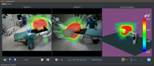
Fig. 4: Augmented Reality visualization of the intensities and of the 3D propagation of scattered radiation for the current C-arm projection provided by our radiation awareness system.
References: Loy Rodas N., 2018. Context-aware radiation protection for the hybrid operating room. PhD Dissertation, University of Strasbourg, France.
We have also included an open-source implementation of the approach from [7] for a real-time human pose estimation in one of the ceiling cameras' view.
The upper-body joints' poses from the people in the room are displayed in 2D over the color image and their current 3D position is also shown in the virtual visualization (see figure 5).
This information is used for providing feedback of the attending persons' radiation exposure.
Indeed,
the human poses' information is applied to segment the attending persons' shapes on the color images.
The obtained foreground masks are then colored according to the simulated radiation intensity at each 3D location to provide feedback about the exposure to the persons' body-parts.
An example is shown in figure 6,
where it can be observed that the clinician standing on the X-ray source's side is more exposed to scattered radiation.
His left arm,
which is positioned directly on the beam's path,
is colored in orange/red.
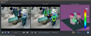
Fig. 5: Clinician tracking using the approach from [7]: the 2D body-joints' positions are overlaid over the color image (left) and the persons' 3D positions are shown in a virtual environment (right).
References: Loy Rodas N., 2018. Context-aware radiation protection for the hybrid operating room. PhD Dissertation, University of Strasbourg, France.
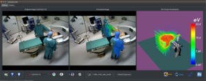
Fig. 6: Augmented Reality visualization of the radiation dose to the clinical staff's body-parts.
References: Loy Rodas N., 2018. Context-aware radiation protection for the hybrid operating room. PhD Dissertation, University of Strasbourg, France.

