ECR 2019 / C-0261
Not all acute pain in the left iliac fossa is diverticulitis.
Congress:
ECR 2019
Poster Number:
C-0261
Type:
Educational Exhibit
Keywords:
Inflammation, Infection, Acute, Surgery, Diagnostic procedure, Ultrasound, CT, Emergency, Abdomen
Authors:
A. B. Barba Arce1, C. González-Carrero Sixto2, J. Azcona Saenz2, J. Crespo del Pozo3, R. Pellón2, A. Cardín Pereda4, E. herrera romero4, E. Marín Diez2; 1Torrelavega, Cantabria/ES, 2Santander/ES, 3Selaya, Cantabria, España/ES, 4santander, cantabria/ES
DOI:
10.26044/ecr2019/C-0261

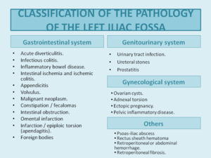
Fig. 1
. References: Department of Radiology, Hospital Universitario Marqués de Valdecilla - Santander/ES")
Fig. 2:
Acute diverticulitis with abscess formation (red arrows).
. References: Department of Radiology, Hospital Universitario Marqués de Valdecilla - Santander/ES")
Fig. 3:
Perforated diverticulitis. Several bubbles of pneumoperitoneum adjacent to the...
. References: Department of Radiology, Hospital Universitario Marqués de Valdecilla - Santander/ES")
Fig. 4:
A 35-year-old man presented with pain in LIF and fever. Upon suspicion of...
. The findings are related to an ischemic colitis. References: Department of Radiology, Hospital Universitario Marqués de Valdecilla - Santander/ES")
Fig. 5:
Patient with pain in LIF: concentric thickening of the descending colon and...
, as well as portal pneumatosis (red arrow). References: Department of Radiology, Hospital Universitario Marqués de Valdecilla - Santander/ES")
Fig. 6:
Case of advanced intestinal ischemia with parietal pneumatosis and thinning...

Fig. 7:
Patient who comes for pain in LIF, before suspicion of diverticulitis is...
. References: Department of Radiology, Hospital Universitario Marqués de Valdecilla - Santander/ES")
Fig. 8:
Sigma volvulus. We can observe the rotation of vessels, meso and sigma in the...
, compatible with neoformation, which causes obstruction of the colon with abundant fecaloid content in the colic frame. Additionally, hepatic metastases were observed (yellow arrow). References: Department of Radiology, Hospital Universitario Marqués de Valdecilla - Santander/ES")
Fig. 9:
48-year-old man who comes for pain in LIF. Stenosing mass in rectosigmoid...

Fig. 10:
Fecaloma.
 with trapped small bowel loop that causes an obstructive condition with dilatation of the loops retrograde. References: Department of Radiology, Hospital Universitario Marqués de Valdecilla - Santander/ES")
Fig. 11:
A left inguinal hernia (red arrow) with trapped small bowel loop that causes an...
 in a 27-year-old girl who was pain in LIF. An ultrasound was performed in the first place and later the surgery service requested to perform a CT scan given the persistence of the pain. We can see the target sign and the pseudokidney sign. References: Department of Radiology, Hospital Universitario Marqués de Valdecilla - Santander/ES")
Fig. 12:
Jejunal-jejunal intussusception (yellow arrow) in a 27-year-old girl who was...
 was observed that also conditioned inflammatory changes in the adjacent musculature. References: Department of Radiology, Hospital Universitario Marqués de Valdecilla - Santander/ES")
Fig. 13:
Woman with pain in FII. Omental infarction (yellow arrow) was observed that...

Fig. 14:
Epiploic appendagitis
. References: Department of Radiology, Hospital Universitario Marqués de Valdecilla - Santander/ES")
Fig. 15:
Lithiasis in left ureteropelvic junction (red arrow).
 caused by lithiasis in the juxtavesical ureter (yellow arrow). In addition, free fluid is observed adjacent to the ureter in relation to rupture of the urinary tract (green star). References: Department of Radiology, Hospital Universitario Marqués de Valdecilla - Santander/ES")
Fig. 16:
Left ureterohydronephrosis (red arrow) caused by lithiasis in the juxtavesical...
. We can see several lithiasis in the calyceal groups and in the juxtavesical ureter (red arrow).
Figure B: Focal nephritis area. References: Department of Radiology, Hospital Universitario Marqués de Valdecilla - Santander/ES")
Fig. 17:
Emphysematous pyelonephritis left. Gas bubbles are observed in the renal...
 by clots, detritus and membranes. In addition, a large left abdominal collection is observed (blue star). The findings were secondary to perforated gangrenous cystitis.
References: Department of Radiology, Hospital Universitario Marqués de Valdecilla - Santander/ES")
Fig. 18:
Patient with pain in the left iliac fossa and fever. Gas bubbles are observed...

Fig. 19:
A 23-year-old girl who presented with abdominal pain and hypotension. In these...

Fig. 20:
15-year-old girl who comes for abdominal pain. It was observed on ultrasound an...

Fig. 21:
12-year-old girl with severe abdominal pain in LIF. An ultrasound was performed...

Fig. 22:
Woman who comes for pain in LIF, we observed a left ovary increased in size...

Fig. 23:
Patient with pain in left iliac iliac fossa. A CT was performed and dilatation...

Fig. 24:
Two cases of patients under treatment with anticoagulants, with spontaneous...

Fig. 25:
Spontaneous anterior rectus hematoma in a patient taking oral anticoagulants....
. References: Department of Radiology, Hospital Universitario Marqués de Valdecilla - Santander/ES")
Fig. 26:
Retroperitoneal hematoma conditioned by the spontaneous rupture of the left...

Fig. 27:
Figure A: Patient with retroperitoneal fibrosis surrounding the aorta.
Figure...
 is observed that displaces the intestinal loops, with fatty densitometric values, suggestive of mesenteric lipoma vs liposarcoma. References: Department of Radiology, Hospital Universitario Marqués de Valdecilla - Santander/ES")
Fig. 28:
Woman who comes for pain in LIF. A large mesenteric mass (blue star) is...

Fig. 29:
The same patient underwent MRI where the homogenous fat component of the mass...
. A large mass was observed with cystic areas of probable left adnexal origin, suggestive of cystadenoma. References: Department of Radiology, Hospital Universitario Marqués de Valdecilla - Santander/ES")
Fig. 30:
35 year old woman with pain in LIF. An ultrasound was performed and a large ...