ECR 2019 / C-2289

Evaluation of Palpable Scrotal Pathology: Diagnostic Imaging Findings at Ultrasound
Congress:
ECR 2019
Poster Number:
C-2289
Type:
Educational Exhibit
Keywords:
Education, Ultrasound-Colour Doppler, Ultrasound, Genital / Reproductive system male, Neoplasia
Authors:
I. D. Oh1, J. Munroe2, G. Sostre2, M. Roda3, P. Mittal4; 1Suwanee, GA/US, 2Augusta, GA/US, 3Jackson, MS/US, 4Decatur, GA/US
DOI:
10.26044/ecr2019/C-2289

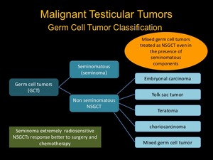
Fig. 5:
Old Classification of Germ Cell Tumor

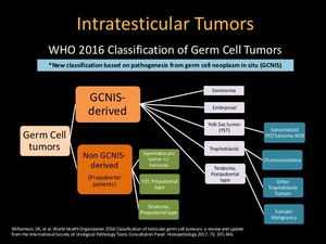
Fig. 6:
2016 WHO Classification of Germ Cell Tumor

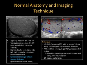
Fig. 7:
Normal Anatomy and Imaging Technique

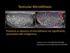
Fig. 8:
Testicular Microlithiasis

Fig. 9:
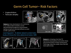
Germ Cell Tumor - Risk Factors

Fig. 10:
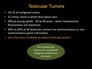
Teseticular Tumor