ECR 2019 / C-2289

Evaluation of Palpable Scrotal Pathology: Diagnostic Imaging Findings at Ultrasound
Congress:
ECR 2019
Poster Number:
C-2289
Type:
Educational Exhibit
Keywords:
Education, Ultrasound-Colour Doppler, Ultrasound, Genital / Reproductive system male, Neoplasia
Authors:
I. D. Oh1, J. Munroe2, G. Sostre2, M. Roda3, P. Mittal4; 1Suwanee, GA/US, 2Augusta, GA/US, 3Jackson, MS/US, 4Decatur, GA/US
DOI:
10.26044/ecr2019/C-2289

Fig. 5:
Old Classification of Germ Cell Tumor

Fig. 6:
2016 WHO Classification of Germ Cell Tumor

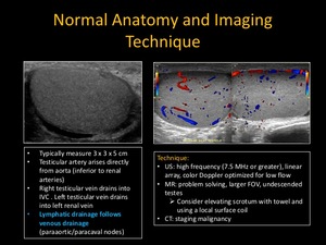
Fig. 7:
Normal Anatomy and Imaging Technique

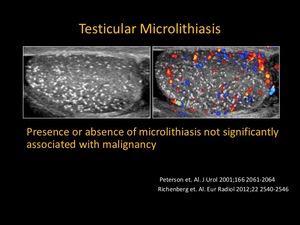
Fig. 8:
Testicular Microlithiasis

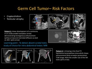
Fig. 9:
Germ Cell Tumor - Risk Factors

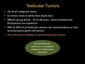
Fig. 10:
Teseticular Tumor
. Testicular tumors: what radiologists need to know—differential diagnosis, staging, and management. Radiographics, 35(2), 400-415.")
Fig. 11:
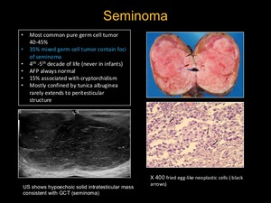
Seminoma
. Testicular tumors: what radiologists need to know—differential diagnosis, staging, and management. Radiographics, 35(2), 400-415.")
Fig. 12:
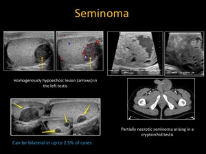
Seminoma

Fig. 13:
Embryonal Cell Carcinoma
. Testicular tumors: what radiologists need to know—differential diagnosis, staging, and management. Radiographics, 35(2), 400-415.")
Fig. 14:
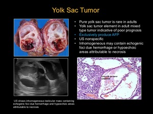
Yolk Sac Tumor

Fig. 15:
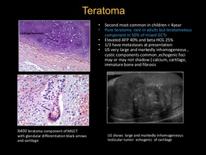
Teratoma

Fig. 16:
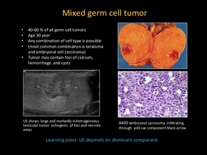
Mixed germ cell tumor

Fig. 17:
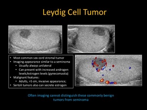
Leydig Cell Tumor

Fig. 18:
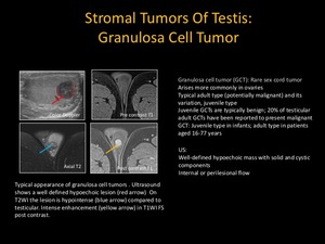
Stromal Tumors of Testis: Granulosa Cell Tumor

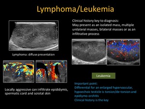
Fig. 20:
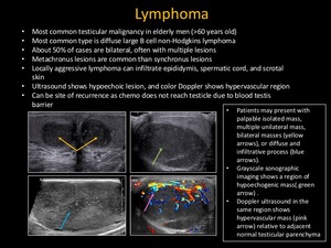
Lymphoma and Leukemia

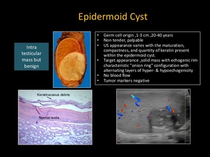
Fig. 21:
Epidermoid Cyst

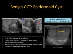
Fig. 22:
Benign GCT: Epidermoid Cyst

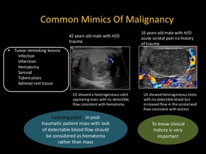
Fig. 23:
Common Mimics of Malignancy

Fig. 24:
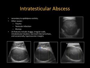
Intratesticular Abscess
. Spectrum of Extratesticular and Testicular Pathologic Conditions at Scrotal MR Imaging. RadioGraphics, 38(3), 806-830.")
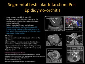
Fig. 25:
Segmental Testicular Infarction : Post Epididymo-orchitis

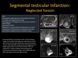
Fig. 26:
Segmental testicular Infarction: Neglected Torsion

Fig. 27:
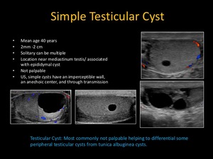
Simple Testicular Cyst

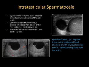
Fig. 28:
Intratesticular Spermatocele

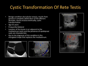
Fig. 29:
Cystic Transformation of Rete Testis
. Spectrum of Extratesticular and Testicular Pathologic Conditions at Scrotal MR Imaging. RadioGraphics, 38(3), 806-830.")
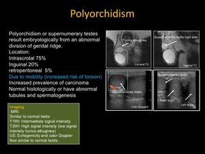
Fig. 30:
Polyorchidism

Fig. 31:
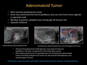
Adenomatoid Tumor

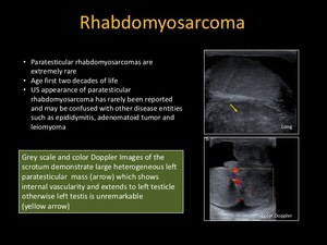
Fig. 32:
Rhabdomyosarcoma

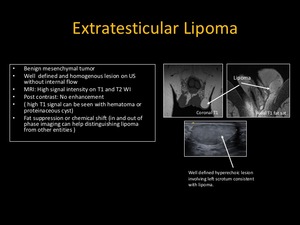
Fig. 33:
Extratesticular Lipoma

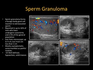
Fig. 34:
Sperm Granuloma
. Spectrum of Extratesticular and Testicular Pathologic Conditions at Scrotal MR Imaging. RadioGraphics, 38(3), 806-830.")
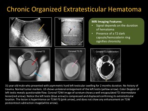
Fig. 35:
Chronic Organized Extratesticular Hematoma
. Spectrum of Extratesticular and Testicular Pathologic Conditions at Scrotal MR Imaging. RadioGraphics, 38(3), 806-830.")
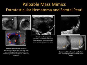
Fig. 37:
Palpable Mass Mimics: Extratesticular Hematoma and Scrotal Pearl

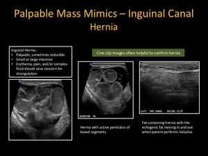
Fig. 38:
Palpable Mass Mimics - Inguinal Canal Hernia

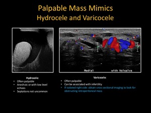
Fig. 39:
Palpable Mass Mimics: Hydrocele and Varicocele

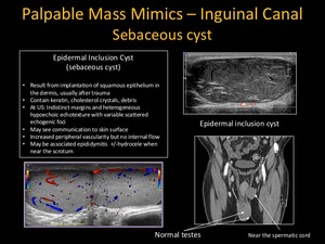
Fig. 40:
Palpable Mass Mimics - Inguinal Canal Sebaceous Cyst

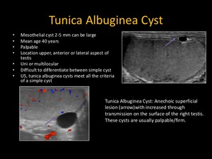
Fig. 36:
Tunica Albuginea Cyst

Fig. 19:
Lymphoma