Keywords:
Haematologic, Respiratory system, Thorax, CT, CT-Angiography, Education, Haematologic diseases
Authors:
A. Bruno, V. Cosi, M. milandri, G. Rasetto, R. Ioppolo, D. Capannelli, C. Sassi, G. Battista; Bologna/IT
DOI:
10.26044/ecr2019/C-2726
Aims and objectives
Introduction
According to the Fleischner Society,
the term reversed halo sign (RHS) defines “a focal,
rounded area of ground-glass opacity surrounded by a more or less complete ring of consolidation” on chest-computed tomography (CT) images (figure 1),
and it resembles the opposite or “reverse” of the ‘‘halo sign’’ which is often found in angioinvasive pulmonary aspergillosis.
It also seems like the ring-shaped coral reefs surrounding a central lagoon,
thus some Authors call it atoll sign (Zompatori et al.
1999),
but the term reversed halo sign is to be preferred,
as stated by the Fleischner society.
The RHS was initially described by Voloudaki et al in 1996 and until 2005-2006 the RHS was considered pathognomonic of organizing pneumonia (OP).
Subsequently,
various Authors demonstrated the presence of this sign in a wide spectrum of diseases,
including infectious and noninfectious conditions as shown in Table 1
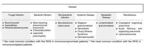
Table 1: Table 1: Spectrum of Diseases with the Reversed Halo Sign (modified from Georgiadou et al. 2011)
It was reported that in neutropenic and/or immunosuppressed patients the presence of the RHS on chest-CT scan should be considered an indication of Invasive Mould Disease (IMD) such as pulmonary mucormycosis (PM) where apparently represents an early phase of the disease. In IMD,
the pulmonary RHS is attributable to pulmonary hemorrhagic infarction with coagulation necrosis of the alveolar septa caused by the invasion and occlusion of peripheral pulmonary arteries by fungal hyphae with a greater amount of hemorrhage at the periphery (the ring-shaped consolidation) than in the center,
where alveolar air remains (the central ground-glass opacity).
It was reported that the RHS is significantly more common in PM than in IPA (P < 0.001) probably due to a greater angioinvasive attitude of Mucorales respect with Aspergillus species.
A recent study by Bourcier et collegues including 27 patients with PM demonstrated that the RHS is more common in neutropenic patients (78%) than in non neutropenic (31%).
To our knowledge,
there are a few studies comparing radiological findings for IPA with those for PM,
and no studies evaluating the specificity of the RHS for PM.
In the present study we retrospectively analyzed the clinical and radiological characteristics of 23 neutropenic onco-hematologic febrile patients with pulmonary lesions showing the RHS compared to a control group of 85 patients without the RHS on chest-CT images (including 44 patients with IMD and 41 without IMD),
to evaluate the specificity of the reversed halo sign and its relations to patient’s immune status.
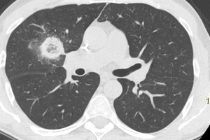
Fig. 1: HRCT finding of pulmonary lesion with the RHS in middle lobe: the nodular lesion consist of a central area of ground glass and a peripheral ring-shaped area of consolidation
