An ischaemic stroke classicaly presents with rapid onset of neurological deficit depending on the area of the brain that is involved.
The vascular territory affected determines the exact symptoms and clinical behaviour of the lesion.
Therefore,
one of the key tasks in establishing the diagnosis of acute ischemic stroke is defining the involved arterial territory.
Thorough knowledge of the vascular territories of the brain are required to confidently diagnose that the diffusion restricted abnormality actually represents an acute arterial stroke.
This further guides the investigations and treatment planning.[1,2,3]
Factors contributing to understanding vascular territories include the anatomy of the intracranial circulation and its normal variants. Basically human brain is supplied by four major arteries,
the paired internal carotids and vertebrals.[2,4]
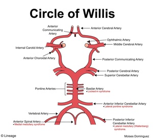
Fig. 22: Circle of Willis
References: Lineage
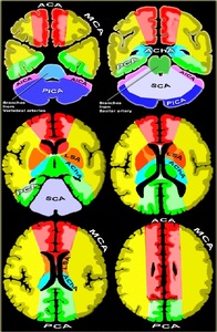
Fig. 23: Vascular territories of the brain
References: http://www.radiologyassistant.com
Internal carotid artery (ICA)
It gives retinal artery and then divides in to the middle and anterior cerebral arteries intracranially.
These are responsible for maintaining the anterior circulation.
Anterior cerebral artery (ACA)
It comprise of four to five segments which supplies the medial frontal lobe,
anterior two-thirds of the medial cerebral hemisphere,
area over the cerebral convexity,
anterior portion of the corpus callosum,
head of caudate,
anteromedial putamen,
globus pallidus and anterior limb of the internal capsule.[1,5]
A1 segment – arise from ICA and extends upto the anterior communicating artery.
Medial lenticulostriate and anterior communicating arteries arise from this segment and supplies the caudate nucleus and the anterior limb of the internal capsule.
A2 segment - Spans from ACom to the level of the genu of corpus callosum.
It bifurcates into the pericallosal and callosomarginal arteries.
The recurrent artery of Heubner arises at the beginning of the A2 segment.
A3 segment- Spans from the genu to the terminal bifurcation.
Pericallosal artery is one of the main terminal branches of the ACA.
A4 segment- Supracallosal arteries are considered as the A4 and A5 segments.
Middle cerebral artery (MCA)
It comprises of four segments.
MCA arises from the internal carotid artery and continues into the lateral sulcus where it typically supplies majority of the lateral surface of the cerebral hemisphere,
including the fronto-temporo-parietal lobes and most of the basal ganglia. Involvement of the basal ganglia indicates a proximal occlusion with lenticulostriate artery occlusion.
Complete occlusion presents as involvement of the the basal ganglia,
insula and fronto-parieto-temporal lobes.[6,7]
M1 segment - Supplies the basal ganglia through lateral lenticulostriate arteries.
M2 segment - Extends anteriorly into the insula to be known as the insular/sylvian segment.
This may bifurcate or trifurcate into segments which extend towards the cerebral cortex.
M3 segment - Extends from the insula towards the cortex.
M4 segments - Also known as corticl segment extend distally on the cortex of the brain.
Vertebral artery
It originates from the subclavian arteries.
Each vertebral artery gives posterior cerebellar artery and then fuse with the contralateral aartery to form the basilar artery.
Basilar artery
It divides into two posterior cerebral arteries which are responsible for maintaining the posterior circulation.
Posterior cerebral artery (PCA)
It comprises of four segments and begins near the union of the posterior communicating artery and basilar artery.
It communicates with the ipsilateral middle cerebral and internal carotid arteries via the posterior communicating artery.
The arterial territory includes inferolateral temporal and occipital lobes,
posterior one third of the brain along the interhemispheric fissure and lower parietal,
occipital,
and temporal lobes.[7]
Perforators and posterior choroidal arteries also supply thalamus,
hypothalamus,
internal capsule posterior limb,
upper midbrain and choroid plexus
P1 segment - Spans from the termination of the basilar artery to the posterior communicating artery.
It give off the posterior thalamoperforator branch.
P2 segment - Starts from the posterior communicating artery and winds around the midbrain.
Many choroidal perforator branches arise from this segment.
P3 segment - It arises within quadrigeminal cistern.
P4 segment - It gives rise to the anterior temporal artery,
posterior temporal artery,
lateral occipital artery,
anterior inferior temporal artery,
middle inferior temporal artery,
posterior inferior temporal artery,
medial occipital artery,
calcarine artery,
parieto-occipital artery and splenial artery.
Superior Cerebellar Artery (SCA)
It arises from the distal basilar artery just below the posterior cerebral artery.
It has prepontine,
ambient and quadrigeminal segments.
It supplies entire tentorial surface of the cerebellar hemispheres,
superior vermis,
dentate nucleus,
most of the cerebellar white matter,
part of mid brain and superior and middle cerebellar peduncles.
Anterior inferior cerebellar artery (AICA)
Each artery arises from the basilar artery at the level of the junction between the medulla oblongata and pons.
It gives off the internal auditory or labyrinthine artery in most cases. AICA supplies the anteroinferior surface of the cerebellum,
the flocculus,
middle cerebellar peduncle and inferolateral portion of the pons.[8]
Posterior Inferior Cerebellar Artery (PICA)
It is the largest branch of the vertebral artery and has four segments.
The anterior medullary segment,
lateral medullary segment,
posterior medullary segment and supratonsillar segment. It supplies the posteroinferior cerebellar hemisphere,
inferior portion of the vermis,
lower part of the medulla and inferior cerebellar peduncles.[8]
Anterior Choroidal artery
It originates from the internal carotid artery between the origin of posterior communicating artery and internal carotid termination.
Rarely it arises from the middle cerebral artery. It is divided into two segments:
Cisternal segment - Starts from its origin until the choroidal fissure and supplies the hippocampus,
optic tract,
lateral geniculate nucleus,
lateral aspect of the thalamus,
posterior limb of internal capsule and lateral aspect of mid brain.
Interventricular segment - Supplies the choroid plexus of the anterior part of the temporal horns.


