Type:
Educational Exhibit
Keywords:
Neuroradiology brain, Oncology, MR-Diffusion/Perfusion, MR, Imaging sequences, Technical aspects, Tissue characterisation, Neoplasia
Authors:
J. Markus, M. Sokolska, R. Jager, H. Hyare; London/UK
DOI:
10.26044/ecr2019/C-3808
Background
Arterial Spin Labelling (ASL) is a completely non-invasive MRI perfusion technique.
The technique utilises train of electromagnetic pulses to label arterial blood water flowing to the brain.
Together with a control image,
acquired without labelling, it produces information on cerebral blood flow.
The use of an endogenous tracer is an attractive pro in the controversial topic of MRI contrast agents.
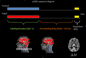
Fig. 1: Using inversion pulses at the labelling plane, inflowing blood water is magnetically labelled. Another, control acquisition is acquired and the difference between the two yields a perfusion weighted image
ASL can produce quantified data,
which is a valuable resource, however this requires offline processing which means time and man power. ASL Perfusion Weighted Images (ASL-PWI) maps can be produced at the time of scanning.
Systems can be programmed to produce these maps with little to no input from the radiographer or other professionals meaning ASL-PWI can be automatically produced and archived with minimal change to exsisting services.
MR perfusion techniques have proven to provide information not available on conventional imaging and can provide information regarding tumour type and grade,
patient prognosis and treament evaluation.
MR perfusion techniques have also been considered as a solution to differentiating between treatment changes and disease progression which is a known pitfall of conventional MRI techniques.
ASL techniques have been utilised in many clinical applications, particularly in imaging brain tumours,
however it remains underused in the UK.

