Keywords:
Experimental, Not applicable, Dosimetric comparison, Physics, Dosimetry, CT, Radioprotection / Radiation dose, Performed at one institution
Authors:
K. Takegami1, H. Hayashi2, T. Asahara1, S. Goto1, E. Tomita2, N. Kimoto2, Y. Kanazawa3, S. Kudomi4; 1Kanazawa, Ishikawa/JP, 2Kanazawa/JP, 3Tokushima/JP, 4Yamaguchi/JP
DOI:
10.26044/ecr2020/C-01393
Methods and materials
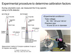
Fig. 2 shows the experimental materials. We measured X-ray spectra in a free-air and at the surface of cylindrical acrylic phantoms (T40027, PTW) having diameters of 16 cm and 32 cm. Acrylic scatterers were then placed in free-air (Fig. 3(a)) and on the phantoms (Fig. 3(b)). Here, volumetric CT scans were performed using a CT scanner (SOMATOM Force, Siemens Healthineers), and Compton-scattered X-rays at 90 degrees [10] were measured using a CdTe spectrometer (type123, EMF Japan). To reduce the pile-up effect and dead time of the CdTe spectrometer, tube current-irradiation time products for each tube voltage of 80-140 kV were flexibly adjusted as shown in Fig. 4. Also, the detector collimation in the CT scanner was set to properly irradiate the acrylic scatterer; 32-detector rows with the size of one pixel being 0.6 mm. In addition, we measured X-ray spectra without the acrylic scatterer to subtract background X-rays.
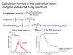
Using the measured X-ray spectra, we determined the CFs of the OSL dosimeter in the following procedures. First, the background X-rays were subtracted from the measured X-ray spectra. Second, the subtracted spectra were corrected by considering a response function of the CdTe spectrometer, which was derived using Monte-Carlo simulation code (an electron gamma shower ver.5 (EGS5)) [11,12]. Next, to derive spectra being incident to the acrylic scatterer, the above-mentioned spectra were corrected for the X-ray energy and the probability of the Compton-scattered X-ray at 90 degrees, which is derived by the Klien-Nishina formula [10]. From the corrected spectra, we derived effective energies based on the evaluation of a half value layer of aluminum and the CFs (Fig. 5), which were calculated from the ratio of kerma and the absorbed dose while taking into consideration the energy dependence of the OSL dosimeter [13]. Finally, we plotted the CFs corresponding to the effective energies, and we evaluated the uncertainty of the CFs.
In order to evaluate the CFs, we additionally performed the Monte-Carlo simulation using the EGS5 code. As shown in Fig. 6, we constructed a small-type OSL dosimeter under previously reported simulation conditions [14], and X-rays of 107 were introduced to the dosimeter. Here, we derived the CFs of monoenergetic X-rays less than 150 keV at every 10 keV and continuous X-rays of 80 kV. The continuous X-rays were derived using Tucker’s formula [15], and an aluminum filter having a thickness of 2.5 mm was added as a total filtration. The CFs and the effective energies were calculated using the same procedure as the analysis of experimental data.


