Keywords:
Experimental, Not applicable, Dosimetric comparison, Physics, Dosimetry, CT, Radioprotection / Radiation dose, Performed at one institution
Authors:
K. Takegami1, H. Hayashi2, T. Asahara1, S. Goto1, E. Tomita2, N. Kimoto2, Y. Kanazawa3, S. Kudomi4; 1Kanazawa, Ishikawa/JP, 2Kanazawa/JP, 3Tokushima/JP, 4Yamaguchi/JP
DOI:
10.26044/ecr2020/C-01393
Purpose
Recently, patients have more opportunities to have CT examinations [1] and the radiation doses to the patients have also increased. It is increasingly important to consider the balance between radiation dose and image quality [2]. In order to evaluate an exposure dose during CT scan, the CT dose index (CTDI) is often used to evaluate the exposure dose of a standardized patient [3] and is built in current CT scanners. Actually, the radiation dose to patients varies depending on the patient size and scanning parameters. It is considered that the CTDI values are not suitable to evaluate the patient dose [3]. In this study, we plan to evaluate individual patient dose by the direct measurement, which can be performed without scan parameter information. We think that the actual measurement is useful as one of the objective evaluations of exposure doses during CT examinations.
Radiation doses to patients have been measured using various radiation detectors such as optically stimulated luminescence (OSL) dosimeters [4], thermo-luminescence dosimeters (TLD) [5], and radiochromic film [6] etc. We focused our attention on the direct measurement of the entrance-surface dose (ESD) to patients using a small-type OSL dosimeter (nanoDot, Landauer, Inc.). To perform the ESD measurement, the OSL dosimeter is placed on the surface of the patient’s skin which is inside the scanning area. For medical imaging, the OSL dosimeters should not interfere with the image. Among various dosimeters, the OSL dosimeter, which is composed of Al2O3:C, has a low detection efficiency, therefore additional artifacts on the medical images were not produced [7]. The dosimeter was found to be suitable to measure the actual dose during a CT examination.
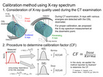
In order to evaluate the exposure dose, it is important to calibrate a dosimeter using a proper method. In the conventional X-ray system, the dose calibration is performed using the relationship between reference air-kerma and/or ESD and the response of the dosimeter. Air-kerma for direct X-rays is measured with an ionization chamber. The ESD is calculated using air-kerma and a back-scatter factor [8] for the contamination of scattered X-rays from the patient. This procedure cannot be applied to the precise ESD measurement during CT examinations because various X-rays are irradiated from all directions (Fig. 1). Recently, Asahara et al. proposed a novel calibration procedure taking into consideration the X-ray quality to measure the actual dose of the patients and medical staffs [9]. As shown in Fig. 1, a calibration factor (CF) can be determined when we know the incident X-ray spectra at the dosimetric point and the energy dependence of the dosimeter. A purpose in the present study is to apply the calibration procedure to determine the CF for the ESD measurement in the CT scanning. The X-ray spectra on cylindrical acrylic phantoms were evaluated based on the measurement of Compton-scattered X-rays at 90 degrees [10].
In order to verify the accuracy of the experimentally determined CFs, we evaluate the CFs using a Monte-Carlo simulation. By comparing the results, we discussed the importance of the calibration procedure taking into consideration the X-ray quality and the uncertainty of the CFs for practical use in clinical situations.


