ECR 2020 / C-04512
High-value multidetector CT angiography for diagnosis of acute mesenteric ischemia.
Congress:
ECR 2020
Poster Number:
C-04512
Type:
Educational Exhibit
Keywords:
Performed at one institution, Not applicable, Retrospective, Acute, Diagnostic procedure, CT-Angiography, Catheter arteriography, Interventional vascular, Gastrointestinal tract, Emergency, Emergency Imaging
Authors:
S. Brugger1, R. M. Piqueras Olmeda1, M. Ballesta2, P. Estelles Lerga1; 1Valencia/ES, 2VALENCIA, Valencia/ES
DOI:
10.26044/ecr2020/C-04512

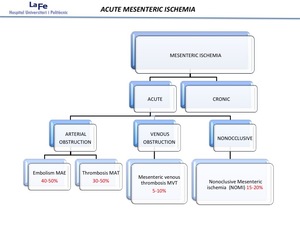
Fig. 1:
Causes of vascular insuffiency.

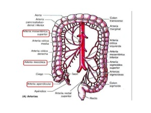
Fig. 2

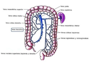
Fig. 3

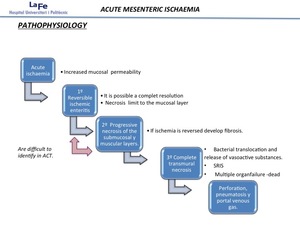
Fig. 4:
Pathophysiology of intestinal ischemia