ECR 2020 / C-05803
Do not forget to take a look at the susceptibility-weighted imaging: Cerebral Amyloid Angiopathy
Congress:
ECR 2020
Poster Number:
C-05803
Type:
Educational Exhibit
Keywords:
Performed at one institution, Not applicable, Pathology, Image verification, Haemorrhage, Surgery, Complications, Biopsy, MR, CT-Angiography, CT, Vascular, Neuroradiology brain, Haematologic, Neuro
Authors:
K. Sotomayor1, A. Bonilla2, M. NEGROTTO3, F. Bustamante1, C. Scelsi1, R. Figueroa1, S. Forseen1, B. Gilbert1, G. Palacios1; 1Augusta, GA/US, 2Lima/PE, 3Montevideo/UY
DOI:
10.26044/ecr2020/C-05803

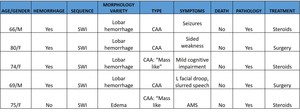
Table 1:
Findings and procedure details.
 Axial T2 (Fig.1B ) Sagittal T2 (Fig. 1C) Coronal T2 (Fig. 1D) FLAIR. Vasogenic edema pattern involving the right anterior frontal lobe.
(Fig. 1E) Coronal T1 (Fig. 1F) Coronal T1+Gd Leptomeningeal enhancement over the right middle and lateral aspect of right inferior frontal gyri. (Fig. 1G) Axial DWI shows no restriction.
References: Department of Radiology and Imaging, Medical College of Georgia, Augusta University/ Georgia- USA 2019.")
Fig. 1:
75-year-old female who recently experienced an alteration in consciousness and...
 (Fig. 2B) (Fig. 2C) (Fig. 2D) axial SWI Minip. Multiples nodular focis of magnetic susceptibility throughout the subcortical white matter of the bilateral cerebral hemispheres.
(Fig. 2E) MRI Spectroscopy. Increased Cho and Myoinositol and decreased NAA
References: Department of Radiology and Imaging, Medical College of Georgia, Augusta University/ Georgia- USA 2019.")
Fig. 2:
The same patient
(Fig. 2A) (Fig. 2B) (Fig. 2C) (Fig. 2D) axial SWI Minip....
 Coronal T2 (Fig. 3B) (Fig. 3C) Axial Flair. Significant interval improvement in the vasogenic edema pattern involving the right anterior frontal lobe. (Fig.3D) Axial T1 +Gd. The previous leptomeningeal enhancement pattern over the right middle and lateral aspect of right inferior frontal gyri has resolved.
References: Department of Radiology and Imaging, Medical College of Georgia, Augusta University/ Georgia- USA 2019.")
Fig. 3:
After MRI of the brain with and without contrast and spectroscopy the lesion...
 (Fig. 4B) (Fig. 4C) (Fig 4D) (Fig 4E) (Fig 4F) Axial SWI Minip. innumerable nodular foci of magnetic susceptibility throughout the subcortical white matter of thebilateral cerebral hemispheres.
References: Department of Radiology and Imaging, Medical College of Georgia, Augusta University/ Georgia- USA 2019.")
Fig. 4:
Follow-up post-treatment
(Fig. 4A) (Fig. 4B) (Fig. 4C) (Fig 4D) (Fig 4E) (Fig...

Fig. 5:
74 year old female with a history of mild cognitive impairment and no other...

Fig. 6:
Congo red: Shows orangeophilia in a few small vessels, with sufficient...
.
Pattern of cerebral injury favoring with multifocal extensive susceptibility signal loss punctate lesions in bilateral cerebral hemispheres with predominant inferior frontal, temporal and left more than right parieto-occipital subcortical distributions, may represent chronic posttraumatic microhemorrhages with associated white matter signal abnormality and parenchymal volume loss pole distributions. References: Department of Radiology and Imaging, Medical College of Georgia, Augusta University/ Georgia- USA 2019.")
Fig. 7:
69 year-old male with L facial droop, slurred speech("code stroke").
Pattern...

Fig. 8:
Congo red: identifies multiple vessels with thickened hyalinized walls that are...