Type:
Educational Exhibit
Keywords:
Performed at one institution, Observational, Retrospective, Haemodynamics / Flow dynamics, Normal variants, Imaging sequences, MR, Veins / Vena cava, Neuroradiology brain, Anatomy, Vascular
Authors:
S. Mahal1, S. Tiwari2, T. Yadav3, P. S. Khera1; 1JODHPUR/IN, 2Jodhpur, Rajasthan /IN, 3110029/IN
DOI:
10.26044/ecr2020/C-07764
Background
The intracranial veins, unlike the systemic veins, do not follow their arterial counterparts and thus differ in their drainage territory from the arteries. Developmentally, the superficial and deep venous system has distinct embryology. Variations in cerebral venous anatomy is a rule rather than an exception. These variations must be kept in mind while evaluating a scan for pathologies like cerebral venous sinus thrombosis(CVT) or assessment of their patency in tumours encasing the venous sinuses1, 2.
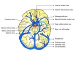
Normal venous anatomy 3: The intracranial venous system has two major components, the dural venous sinuses and the cerebral veins.
Dural Venous Sinuses:
· Endothelium-lined channels, contained between the outer (periosteal) and inner (meningeal) dural layers.
· Subdivided into an anteroinferior group and a posterosuperior group.
· The posterosuperior group is the more prominent, consisting of the superior sagittal sinus (SSS), inferior sagittal sinus (ISS), straight sinus (SS), sinus confluence (torcula herophili), transverse sinuses (TSs), sigmoid sinuses, and jugular bulbs.
· The anteroinferior group consists of the cavernous sinus (CS), superior and inferior petrosal sinuses (SPSs, IPSs), clival venous plexus (CVP), and sphenoparietal sinus (SphPS).
· Bilateral cerebral hemispheres are drained via multiple cortical veins, few of them being prominent and named. The vein of Trolard, vein of Labbé, and superficial middle cerebral veins vary in size, maintaining a reciprocal relationship with each other. If one or two are dominant, the third anastomotic vein is usually hypoplastic or absent.


