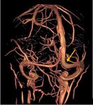
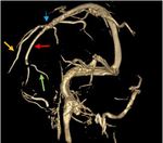
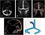
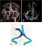
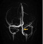
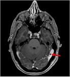
We have reviewed phase-contrast venography and post-contrast 3D –T1 spoiled gradient echo images of 60 patients who came to our institute with a wide spectrum of neurological symptoms. We have described a spectrum of normal as well as variant venous anatomy along with common pitfalls of venous sinus imaging.
Anomalies:
1. Anomalies of the transverse sinus (Fig. 6):
· Most common venous sinus to have anatomic variation, unilateral hypoplasia being the most common variation3.
· Right-sided hypoplasia is more common in females.
· Left-sided hypoplasia equally seen in both males and females4.
2. Anomalies of the superior sagittal sinus (Fig. 7-9):
· Not very common.
· Most frequently reported variation is preferential drainage into one of the TSs, rather than draining at the torcula.
· Segmental or complete duplication of the SSS are associated with occipito-parietal meningoencephaloceles and an anomalous course of the straight sinus5, 6.
Based on the existing literature7, rostral SSS dispositions are classified as following:
a) A normal rostral SSS with cortical veins from both frontal lobes draining into it: most common variety.
b) Duplication of the rostral SSS: two individual channels (hemi-SSS) that have not united at the midline.
c) Completely hypoplastic rostral SSS with compensatory drainage through bilateral large superior frontal veins. This is due to failure of formation of the rostral segment of the sagittal plexus.
d) Unilateral hypoplastic rostral SSS with ipsilateral large superior frontal vein: a tubular parasagittal frontal cortical vein courses on the surface of the brain, rather than the flattened appearance of a dural sinus. It joins the middle third of the SSS near or at the coronal suture. A contralateral small midline venous channel is present which is directly continuous with the middle third of the SSS.
3. Variant anatomy of the torcula herophili 8 (Fig. 10-14): Normally the SSS meets the left and right TS along with the SS at the midline to form a confluence. The group in which this pattern is followed is defined as true confluence (Type I). The group in which three of four sinuses had a union was considered to be partial confluence(Type II and III).
4. Persistent falcine sinus (Fig. 15): It is a normal structure located in the falx cerebri, which involutes after birth. When the SS is absent or rudimentary, the falcine sinus can be recanalized to enable venous drainage in response to the atretic SS and drains into the SSS.
5. Occipital sinus (Fig. 16):
- Originates from the primitive torcular plexus.
- Smallest of the venous sinuses, lies on the inner surface of the occipital bone.
- Drains postero-superiorly at the torcula.
- It important in conditions requiring a posterior fossa craniotomy, as the sinus may be large or in some cases off-midline which makes it amenable to injury9.
6. Brain herniating into arachnoid granulation (Fig. 17):
- Herniation of brain parenchyma along with surrounding CSF into the venous sinus, arachnoid granulation or calvaria, having a prevalence of 0.32%.
- Seen more frequently in postero-inferior parts10.
- More common in females and are thought to be caused by raised intracranial tension.
- To distinguish it from encephalocele, the continuity of the external table of the calvarium is to be looked for. The presence of pre-existing arachnoid granulations, typically giant ones facilitates herniation into venous sinus or adjacent calvarium11-13.
- Rare entity, should be considered in the differential diagnosis of encephaloceles.
Pitfalls in imaging techniques:
A. Arachnoid granulations(Fig. 18, 19):
- Structures protruding into the lumen of the dural sinus: potential to be mistaken as thrombosis.
- Some knowledge regarding the usual and common location is helpful to avoid the pitfall.
- The common locations are TS, particularly the dominant sinus, followed by SSS and the SS.
- Display central signal intensity similar to CSF and appear as focal filling defects with a characteristic anatomic distribution14.
B. Flow-gaps non-contrast venography: TOF MR Venography(MRV) or phase contrast Venography are methods used nowadays for the diagnosis of CVT.
- Venous flow in the plane of image acquisition produce saturation and thus cause drop of venous signal at TOF MRV - source of errors in diagnosis.
- Small-vessel visualization is also superior at CE-MRV, compared with that at TOF MRV15.


