Purpose
Radiological reporting in multiple sclerosis (MS) and other neurological conditions is an important tool that facilitates communication between radiologist and neurologist. In this work we assess the added value of automatically prepopulated radiology reporting templatesfor MS follow-upbased on the automated icobrain software.
Methods and materials
Longitudinal MRI acquisitions approximately 1 year apart were collected from 25 multiple sclerosis patients. The icobrain software [1] was used to compute lesion load (including new and enlarging lesions) and brain volumetry (including annualized atrophy). These findings were automatically inserted in a radiology reporting template.
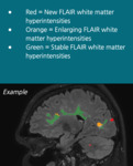
Each MRI dataset, which included images from two time points (TP1 and TP2, see figure 1), was presented in random order to an experienced radiologist twice: once without and once with automatically generated report. In the latter case, besides color-coded...
Results
With respect to timing, the median time for conventional radiological reporting was 7min17s (interquartile range 4min40s), while the median computer-aided reporting time was 4min37s (interquartile range 3min35s), with the latter significantly faster (paired t-test and Wilcoxon signed-rank test p
Conclusion
Computer-aided radiological reportinghas been found to be faster than conventional reporting, with approximately 8 conventional reports per hour versus 13 computer-aided reports per hour.
Stable patients identified by the computer-aided technique were also deemed stable by conventional radiological reading, but the software also reported cases of subtle cerebral atrophy.
We conclude that computer-aided radiological reports are more efficientand bring objectivity to conventional reporting. Standardization of radiologic reporting and incorporation of brain atrophy are steps toward improvement in the clinical management of MS patients.
Personal information and conflict of interest
D. Sima; Leuven/BE - Employee at icometrix D. Smeets; Leuven/BE - Employee at icometrix W. van Hecke; Icometrix, BELGIUM/BE - CEO at icometrix T. Vande Vyvere; Leuven, BE/BE - Employee at icometrix G. Wilms; Leuven/BE - Consultant at icometrix
References
[1] Jain S, Sima DM, Ribbens A, Cambron M, Maertens A, Van Hecke W, et al. Automatic segmentation and volumetry of multiple sclerosis brain lesions from MR images. NeuroImage Clinical 2015; 8: 367-75.


