Keywords:
Performed at one institution, Observational, Retrospective, Multidisciplinary cancer care, Surgery, Removal, Puncture, Percutaneous, MR, CT, Musculoskeletal bone, Interventional non-vascular, Musculoskeletal
Authors:
J. Igrec, I. Brcic, M. Bergovec, R. Igrec, M. A. smolle, B. Liegl-Atzwanger, M. Fuchsjäger, A. Leithner, R. H. Portugaller; Graz/AT
DOI:
10.26044/ecr2020/C-12119
Results
CT was performed in 97 patients, MRI in 106 patients. Table 1
The lesions were most frequently located in the tibia (n=35; 30.4%), the femur (n=34; 29.6%), the humerus (n=9; 7.8%), and the radius (n=5; 4.3%). The rest of the lesions (n=44, 37.6%) were located in hands, sacrum, spine, feet, scapula, fibula, patella, ulna and acetabulum. Lesions were located cortically in 81% of the cases. In 1 patient the lesions were multifocal. Table 2 Table 3 The mean diameter of the nidus was 9.1 ± 5.4 mm (range 2 – 25 mm). A delay in the diagnosis was 11.0 ± 12.8 months.
Due to clinical challenges in the diagnosis of OO, additional information was gathered regarding athletes in our study group (n=7). The mean age of the patients in this group was 22.6 ± 17.2 years, the size of the nidus 9.1 ± 5.7 mm and delay in the diagnosis of 20.7 ± 11.1 months Fig. 8
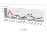
A total of 109 patients underwent treatment at our institution: 55 were treated surgically, and 54 with RFA. Fig. 9
The primary clinical success rate in our study in RFA and surgically treated patients was 88.9%, and 92.7%, respectively. The secondary clinical success was in both groups 100%. In most of the patients, after the tumor recurrence, the same type of procedure was performed. In 2 patients, RFA treatment was initially performed followed by chirurgical excision after the new onset of clinical symptoms and radiologically confirmed tumor recurrence.
In operation specimens (n=44) the diagnosis of OO was histologically confirmed in 100%.


