ECR 2020 / C-13948
Cardiac Fibroma: is T1 mapping “the new tool” for a correct diagnosis?
Congress:
ECR 2020
Poster Number:
C-13948
Type:
Educational Exhibit
Keywords:
Cardiac, Contrast agents, MR, Education, Tissue characterisation, Retrospective, Observational, Performed at one institution
Authors:
S. Pradella1, M. Letteriello1, C. De Amicis1, M. Acquafresca1, E. Bertelli1, G. Grazzini1, V. Miele2; 1Florence/IT, 2FIRENZE/IT
DOI:
10.26044/ecr2020/C-13948
")
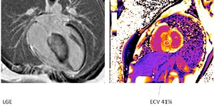
Fig. 7:
Fig. 7: the hyperenhancement of the fibroma on LGE reflected an increased...

Fig. 8:
Fig. 8: ECV is markedly increased in fibromas: in this patient is of 90% due...