Purpose
In vivo imaging and preoperative precise mapping of erectile nerves such as cavernous (CN) can help to improve their preservation during pelvic surgeries [1] such as prostate cancer surgery and thus help in preserving patient’s erectile function [2-4]. Such mapping is particularly challenging since individual CN are microscopic in size (100–400μm) and their number, topology, and location (ventral, dorsal, or lateral) can significantly differ from one patient to another [5,6].A variety of techniques have been developed to identify CN and study their anatomy and physiology,...
Methods and materials
Our methodology includes 3 steps:
(1) MRI scanning of pelvis with four sequences: T2_TSE (AX, COR, SAG),
окT2 mapping (AX), T2_space ZOOMit (AX), T2_space_ SPAIR;
(2) image postprocessing to differentiate and map CN and another significant structures;
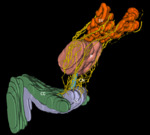
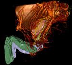
(3) 3D reconstruction of the differentiated nerves.
We started by finetuning these methodology on three volunteers and conducted 12 MRI studies using 3 Siemens MAGNETOM MRI scanners: Aera48 1.5T with two coils Body18, then Skyra48 3T with coil Body60 and finally using Prisma64 3T with coils Spine+Body18. Then,...
Results
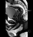
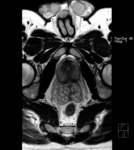
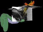
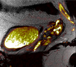
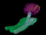
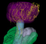
The possibility and accuracy of anatomical visualization of CN on clinical MRI scanners with different magnetic induction was evaluated. As the result of 24 MRI studies, we obtained high resolution images (Fig.1-2) with slice resolution 0.29 х 0.29 х 2.5 mm (real, without interpolation). Subsequently we mapped the variety of nerve fibers 0,2-0,7 mm in diameter spreading from prostate area and then down towards the crus of penis, penetrating the corpus cavernosum and passing inside. We regarded these nerve fibers as CN. CN visualization accuracy...
Conclusion
Our results on humans are among the first on MRI visualization of pelvic plexus and CN with further verification. Thanks to the results of our histological studies our methodology showed high sensitivity and specificity for CN mapping.
Note that MRI Diffusion Tensor Imaging (DTI) can also be used for periprostatic nerves visualization [7], but as demonstrated in [8] visualization of pelvic nerves with DTI is of low specificity. As the next steps we plan to improve our methodology, test developed protocols on advanced MRI 3T...
Personal information and conflict of interest
I. Pyatnitskiy; Moscow/RU - nothing to disclose O. A. Vorontsov; Moscow/RU - nothing to disclose R. Ovchinnikov; Moscow/RU - nothing to disclose V. Mitrokhin; Moscow/RU - nothing to disclose R. Marishin; Moscow/RU - nothing to disclose E. Kharlamov; Oslo/NO - nothing to disclose B. Aleksandrov; Moscow/RU - nothing to disclose S. Droupy; Nîmes/FR - nothing to disclose V. Medvedev; Krasnodar/RU - nothing to disclose
References
[1] Bertrand, M. M. et al.(2014). MRI-based 3D pelvic autonomous innervation: a first step towards image-guided pelvic surgery. European radiology, 24(8),1989-1997.
[2] Fried, N. M., et al. (2015). Novel methods for mapping the cavernous nerves during radical prostatectomy. Nature Reviews Urology, 12(8),451.
[3] Chen, H., et al. "Correlation between the prostate size and location of the neurovascular bundles." J. Endourol 27 (2013): A34-A35.
[4] Haglind, Eva, et al. "Urinary incontinence and erectile dysfunction after robotic versus open radical prostatectomy: a prospective, controlled, nonrandomised trial." European...

