Type:
Educational Exhibit
Keywords:
Neuroradiology brain, MR, Education, Blood
Authors:
S. Mahal, S. Tiwari, T. Yadav, P. K. Garg, P. S. Khera, B. Sureka
DOI:
10.26044/ecr2022/C-15127
Background
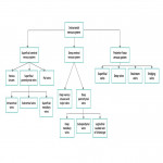
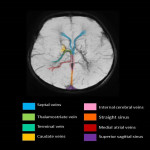
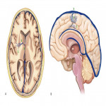
A. Relevant cerebral venous anatomy1 : The intracranial venous system is classified as the (1) superficial cerebral venous system, (2) deep cerebral venous system and (3) posterior fossa venous system (Fig 1,2)
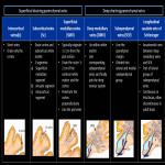
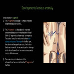
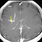
- The superficial cerebral venous system: consists of venous sinuses along with pial veins and the superficial parenchymal veins. The pial and the superficial parenchymal veins drains most of the cerebral cortex and subcortical white matter. The superficial parenchymal veins namely the intracortical, subcortical and superficial medullary veins derive their respective names from their origin and course. (Fig 3)
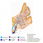
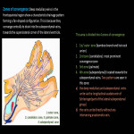
- The deep parenchymal veins comprise of deep medullary veins, subependymal veins along with the longitudinal vein of Schlesinger. The deep medullary veins originate 1-2 cm deep to the pial surface and course centripetally to drain into the corresponding subependymal veins and eventually into the deep venous system (Fig 4,5).
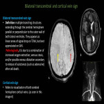
Transcerebral veins : extend from the pial surface, draining into the deep venous system. They penetrate through the cerebral white matter without forming the zones of convergence.
Anastomotic medullary veins : small twigs bridging the superficial and deep medullary veins.
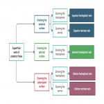
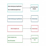
- Posterior fossa veins2: These divided into four groups: superficial, deep, brainstem, and bridging veins. (Fig 8)
- Superficial veins: These are classified on the basis of the surface of the cerebellum they drain and whether they drain the hemispheres or the vermis (Fig 9)
- Deep veins: These veins course in the three deep fissures and on the three cerebellar peduncles.
-Veins of cerebellomesencephalic fissure
-Veins of cerebellomedullary fissure
-Veins of cerebellopontine fissure
-Vein of superior cerebellar peduncle
-Vein of middle cerebellar peduncle
-Vein of inferior cerebellar peduncle
-Deep tonsilar veins
- Veins of the brainstem: They are classified based on 3 characteristics-
-Part of the brainstem drained by them- mesencephalic/pons/medulla
-Surface of the brain drained- median anterior/ lateral anterior
-Direction of their course- transverse/longitudinal.
- Bridging veins: Connect the cortical veins with the dural venous sinuses. Divided into 4 groups:
a. Superior sagittal group
b. Sphenoidal group
c. Tentorial group
d. Falcine group
B. Correlation between the deep medullary veins and white matter tracts1:
Research points to correlation between formation of convergence zones with the crossing nerve fiber tracts in the white matter. In the frontoparietal region, the second zone corresponds to the areas between the association fibers and projection fibers of the corona radiata. Similarly, the third zone fits between the projection fibers of the corona radiata and is called the superior occipitofrontal fasciculus.


