Breast cancer is the most common cancer among women, except for skin cancers. (1) It remains a second leading cause of cancer-related death in women, surpassed by lung cancer only.
About one in eight (13%) women in the USA will develop invasive breast cancer during their lifetime (1). According to American Cancer Society, about 281,550 new cases of invasive breast cancer is estimated in women in 2021, and 49,290 new cases of carcinoma in situ expected to be diagnosed (carcinoma in situ is noninvasive and is the earliest form of breast cancer) (1).
Imaging modalities such as mammography, ultrasonography and magnetic resonance imaging (MRI) are routinely used to identify and characterize breast lesions. (2, 3) Mammography, for all its limits, is still the only test proven to decrease mortality in multiple, randomized, controlled trials and through experience with population-based screening. (4) There are known limitations of mammography, especially in women with extremely dense breast tissue and inconsistent results when this technique is utilized in detecting small, non-palpable lesions. (5)
MRI is recognized as a supplement to mammography in the visualization and characterization of breast lesions and over the last decade, MRI was widely adopted for assessment of breast disease, especially in women with high familial risk. (5,6) MRI cannot visualize calcium deposits that typically surround ductal carcinoma in situ (DCIS) lesions. Mammography can detect these calcium deposits and architectural distortions. (5,6)
Conventional radiological imaging modalities (e.g. mammography, ultrasonography, and MRI) were used to characterize lesions of 84 women in a cross sectional study.
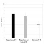
Consent was obtained for the extra experimental quantitative MRI sequence, which was performed at the end of the routine MRI examinations. Subjects were scanned with the mixed-TSE pulse sequence, which is multispectral in T1 and T2, therefore, affords maps of T1/T2 for quantitative assessment of lesions on MRI. The qMRI research sequence and imaging processing were described in details previously. (7)
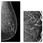
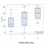
Analysis of microcalcifications, which were detected on mammography, was performed in correlation with histopathology findings (e.g. stage of tumor, size of lesion, estrogen, progesterone, Her2/neu receptor status).


