A) CINEMATIC RENDERING TECHNIQUE (CRT)
Cinematic rendering technique (CRT) transforms standard CT and MRI data into photorealistic 3D images, by precisely simulating light propagation and interaction. Unlike volume rendering technique (VRT), cinematic rendering technique (CRT) enhances depth perception in 3D reconstructions, crucial for through-plane views.
It utilizes high dynamic range rendering maps for natural lighting, resulting in a visually appealing and lifelike image, prioritizing depth and shape perception over spatial resolution [1].
CRT could be valuable for presurgical and preinterventional planning where more advanced visualization techniques are not utilized and could become a standard visualization tool for help in various surgical fields, such as orthopedics, neurosurgery, genitourinary surgery, etc. CRT allows surgeons to visualize anatomical details from various perspectives, closely resembling what they would encounter in the operating room. The increased realism may help mitigate the impact of unexpected findings and anatomical variations during surgery [1].
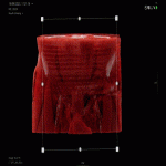
Advanced visualization laboratories are emerging globally. Yet, radiology departments lacking one can use the CRT as a virtual autopsy. By adjusting settings, radiologists can emphasize pathology, aiding surgeons in better treatment planning (Figure 1).
CRT provides a lifelike portrayal of anatomical features, offering improved depth and structure delineation. In instances where 3D printing is unavailable, it can serve as a viable alternative [1, 2].
B) EXTENDED REALITY (XR)
Extended reality (XR), facilitated by computer technologies, merges real and virtual environments, enabling interactive human-machine experiences. Virtual reality (VR) and augmented reality (AR) lead the pack as the most popular technologies within this domain [2,3].
VR involves using computer technology to construct an entirely immersive simulation of a 3D environment. Users can interact with this environment in a realistic, physical manner, often facilitated by hand controllers. In contrast, AR employs computer technology to overlay virtual 3D objects onto the real environment [2,3].
3D printing, augmented reality (AR), and virtual reality (VR) are utilized as 3D imaging techniques to display anatomical models. The process involves converting original stacks of 2D radiological images (i.e., DICOM data) into a 3D volume, comprising image acquisition, segmentation, and computer-aided design/modeling (CAD/CAM). The manipulated data is then optimized for the chosen visualization method — 3D printing, AR, or VR.
These 3D models, derived from DICOM data, simplify the understanding of intricate diseases. By elucidating complex anatomical structures and highlighting pathology, clinicians can effectively plan surgical procedures, thereby improving patient outcomes [3].
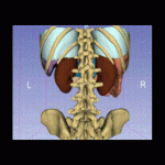
So, the next step is immersing surgeons in a virtual 3D space with VR goggles. CAD 3D models are previously segmented and generated using software and imported into visualization applications enabling CAD 3D model manipulation using controllers as seen in Figure 2.
3) 3D PRINTING AND MODELING
CT images are frequently preferred for 3D printing due to their versatility and ease of postprocessing. The notable contrast, signal-to-noise ratio, and spatial resolution improve the distinction of structures and reduce potential issues like partial volume effects that might constrain 3D printing [4].
Creating anatomically precise 3D models requires meticulous planning, precise acquisition of DICOM data, specialized software for segmentation, and computer-aided design (CAD) for manipulating 3D components, and the expertise of skilled operators [3,4].
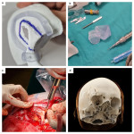
DICOM images are processed using software for segmentation. A radiologist validates the segmentation, and the ordering provider approves the digital model for intended use. The CAD 3D models can be additionally modeled in terms of correcting errors, adding identification marks, etc., using appropriate 3D modeling software. The digital 3D model is then converted using "slicer" software into commands for the 3D printer to produce the physical model [5].
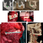
3D printing offers top visual and tactile anatomy experiences. Combined with 3D modeling, it allows creating patient-specific surgical guides and grafts, as seen in Figures 3 and 4 [6,7].
Additionally, open-source technologies and free software are utilized in alternative approaches for mandibular reconstruction planning, providing cost-effective options compared to expensive proprietary software solutions [6].
Preoperative 3D planning and cutting guides improve surgical precision, resulting in better outcomes. Without guides, osteotomies could be less accurate, leading to the need for additional manipulation, such as burring, and slower, less effective healing with increased space between the graft and the healthy mandible [7].
Before the integration of 3D printing, surgical decisions for resection and reconstruction relied on surgeons' experience, often determined intraoperatively. The introduction of 3D printing allowed for improved preoperative planning, providing surgeons with a clear understanding of tumor resection extent, fibular bony fragment size and shape, and osteotomies fitting. This enhances precision and efficiency in restoring facial contour after tumor resection, especially in deciding osteotomies, which would otherwise be done in the operating room without cutting guides, making the process less precise and more time-consuming [6].
Effective utilization of medical 3D printing to improve patient care requires strong interdisciplinary communication, collaboration, knowledge sharing, and a deep understanding of technological advancements. The potential applications of 3D printing in the medical field are boundless and constrained only by one's creative imagination [2,4]."


