Given the high incidence and burden of meniscal related knee pain,
it is crucial that radiologists give accurate and timely diagnosis of meniscal pathology on imaging.
This will aid clinicians in deciding whether conservative treatment or surgical exploration is needed.
Meniscal damage is directly linked with early degenerative osteoarthritis,
features of which are usually present on imaging.
Succinctly relaying these findings to the referrers,
especially to primary care practitioners who may not be familiar with certain terminology is as important as the diagnosis itself.
Whilst arthroscopy remains the gold standard,
MRI demonstrates high sensitivity and specificity in detecting meniscal tears. The aim of this pictorial update is to review and evaluate MRI parameters and protocols that optimise meniscal imaging,
recap crucial meniscal anatomy including variants and discuss key pitfalls that should be avoided.
Introduction
The medial and lateral menisci lie between the tibial plateau and femoral condyle.
They are C-shaped fibro-cartilaginous structures Fig. 1 .
The menisci have several functions as described below:
- to act as shock absorbers
- deepen the contact surface between the femoral condyles and tibial plateau
- act as a stabilisation mechanism
- disperse axial load
- assist in joint lubrication
- aid in proprioceptive mechanisms
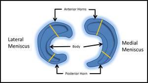
Fig. 1: Image showing the C shaped orientation of the menisci. Note the larger calibre of the medial meniscus in comparison to the lateral. For both, the anterior horn is also larger than the posterior horn.
Tears of the meniscus usually result as a consequence of an acute trauma or secondary to progressive degeneration of interstitial tears to the meniscal surface.
Clinical examination of patients with a meniscal tear usually demonstrates symptoms of joint line tenderness,
locking of the joint and a positive McMurrays stress test.
Tears most commonly occur in the posterior horn of the medial meniscus.
Anterior horn meniscal tears are often over reported and may not always represent a significant tear of concern.
Tears that are asymptomatic are usually oblique or horizontal degenerative type tears.
It is rare to have a complex displaced degenerative tear.


