A consecutive series of patients with isolated lateral malleolar fracture admitted to the emergency room were prospectively enrolled in the period between February 10,
2016,
and November 30,
2017 (21 months).
Patients with skeletal maturity,
lateral malleolus fractures with suspicion of deltoid ligament injury or medial malleolar avulsion up to 5 mm,
lateral malleolus fracture with >2 mm shortening or rotational deviation were included in the study.
Those with previous trauma at the malleolar level,
isolated fracture of the medial malleolus,
posterior or without deltoid ligament injury,
those operated by other colleagues or with more than 15 days after the acute traumatic event,
and those transferred to another hospital were excluded.
With these a longitudinal observational study,
comparative prospective cohort was idealized,
using the gravitational stress radiography as the "gold standard" to determine the state of the deltoid ligament of each of the individuals in the cohort population.
The sample was divided into two groups: group I (Manual External Rotation Stress - MERS) with malleolar fracture associated with injury of the deltoid ligament (exposed patients) or without injury of the deltoid ligament (unexposed patients) and group II (Gravity Stress - GS) with malleolar fracture associated with deltoid ligament injury (exposed patients) or without injury to the deltoid ligament (unexposed patients).
The size of the medial clear space was centered at <4 mm,
or ≥4 mm (4-4.9 mm) and ≥5 mm,
thus allowing the separation of individuals with or without lesion of the deltoid ligaments.
- The MERS test was performed in the operating room under anesthesia,
with the foot in dorsiflexion and 15º of internal rotation [17],
applying external rotation force and visualized in Philips image intensifier.
[Fig.
3]
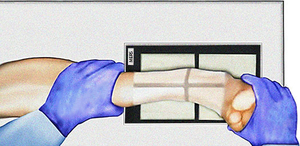
Fig. 2: Manual External Rotation Stress radiograph (MERS)
References: Created by João Oliveira based on original by Dr. Nabil Ebraheim
- GS test was performed with the patient in lateral decubitus,
with a cushion placed laterally and proximal to the injured ankle.
[Fig.
2]
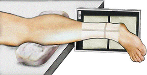
Fig. 3: Gravity Stress radiograph (GS)
References: Created by João Oliveira based on original by Dr. Nabil Ebraheim
In order to avoid bias in the analysis,
the tests were exclusively carried out by the author of the research,
although their interpretation was carried out by independent evaluators blinded to the result between observers.
Similar reading criteria were used for both tests,
measuring the medial clear space between the medial border of the talus and the lateral border of the medial malleolus in millimeters,
drawing a line perpendicular to 5 mm from the upper border of the medial articular surface [Figures 5,6],
similar to what advocated by Travis Motley [14] and Rungprai et al. [18].
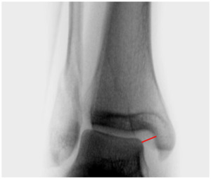
Fig. 4: Medial clear space measurement in MERS x-ray test.
References: Centro Hospitalar Lisboa Ocidental - Lisboa/PT
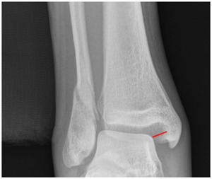
Fig. 5: Medial clear space measurement in GS x-ray test.
References: Centro Hospitalar Lisboa Ocidental - Lisboa/PT
In order to evaluate the intraobserver and interobserver reliability of the measurement of the medial clear space,
the numbers in which the readings of both observers were agreed were added then divided by the total number of measurements read and multiplied by 100. The Kappa value was calculated by the equation:
(% agreement observed) - (% agreement expected just by chance)
______________________________________________________
100% - ( % agreement expected just by chance)
The descriptive statistical analysis of the data was done using the SPSS® v23.
Categorical data was presented as percentages, while continuous variables were characterized using measures of central tendency and dispersion.
The Chi-Square test was used to evaluate the association between two variables,
and the Mann-Whitney test to evaluate the significance,
with a significance level of p<0.05.
As a way of evaluating the linear relationship between the two tests,
the Pearson correlation coefficient was used.
