Aims and objectives
Opto-acoustic (OA) Imagio® (Seno Medical Instruments,
San Antonio,
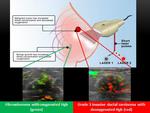
TX) is a novel investigational device (Figure 1) with functional modality that integrates laser optics with ultrasound in an effort to improve the performance of currently available breast imaging modalities without the use of IV contrast or ionizing radiation.
A fusion of anatomic and functional modalities,
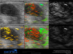
OA imaging provides both conventional B-mode images and co-registered real time color maps that demonstrate tumor vascularity and the relative amount of hemoglobin oxygenation within and around breast tumors.
The technology...
Methods and materials
Study Design
After initial clinical validation,
a Health Insurance Portability and Accountability Act (HIPAA)-compliant institutional review board (IRB)-approved Feasibility Study of OA imaging was undertaken.
155 patients with solid breast masses assessed as BI-RADS 3,
BI-RADS 4,
or BI-RADS 5 on conventional diagnostic ultrasound imaging were enrolled with informed consent. All of these patients were subsequently scanned with OA imaging at one of two IRB-approved sites.
79 lesions were biopsied yielding 39 benign and 34 malignant results,
with 6 lesions excluded,
for a final study...
Results
The total internal OA score,
calculated as the sum of the hemoglobin score,
vessel score,
and blush score within the tumor interior,
correlated positively with histopathologic measures of tumor grade,
though the correlations did not achieve statistical significance (Figure 4).
In contrast,
the total external OA score,
calculated as the sum of the boundary zone and peripheral zone scores,
demonstrated an inverse relationship with histopathologic measures of tumor grade (Figure 5),
which achieved statistical significance for SBR-TOTAL,
SBR-N,
and SBR-M,
though not for SBR-T.
High-grade...
Conclusion
OA Imaging – Pathology Correlation
Opto-acoustic imaging is a new technology that may be able to demonstrate anatomic tumor morphology and provide functional information about tumor vascularity and deoxygenation without the need for contrast injection or ionizing radiation.
OA color maps demonstrate an increased total amount of hemoglobin as well as greater deoxygenation of hemoglobin within and/or surrounding malignant breast tumors.
The distribution of these OA features within the tumor interior versus the external boundary zone and periphery correlates with the histologic grade of the...
References
1. Folkman J.
Tumor angiogenesis: therapeutic implications.
N.
Engl.
J.
Med.
1971;285:1182–86
2. Folkman J.
Clinical applications of research on angiogenesis.
N.
Engl.
J.
Med. 1995;333:1757-1763.
3.
Mahmoud SM,
Paish EC,
Powe DG,
et al.
Tumor-infiltrating CD8+ lymphocytes predict clinical outcome in breast cancer.
J Clin Oncol 2011;29(15):1949-1955.
4.
Berg WB et al.
Shear-Wave Elastography Improves the Specificity of Breast US: The BE1 Multinational Study of 939 Masses.
Radiology.
2012;262(2): 435-449
5. Yoon JH et al.
Shear-wave elastography in the diagnosis of solid breast masses:...


