Aims and objectives
CT plays a very important role in the imaging of oncology patients. However,
the increasing use of CT in the last few decades has raised concerns regarding the risk of cancer from CT imaging.
CT radiation dose is particularly important in oncology patients as they often require serial surveillance scans to monitor for the effectiveness of treatment,
disease progression or recurrence.
The aim of this study was to evaluate if patient CT radiation dose can be reduced without compromising image quality by using a single...
Methods and materials
Seventy oncology patients with previous conventional contrast enhanced (CE) CT CAP referred for restaging or surveillance CE CT CAP were imaged using the proposed protocol of two contrast injections and single-pass acquisition.
The patients were all adult outpatients referred for restaging or surveillance scans.
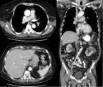
The conventional protocol at our institution is acquired in two segments (figure 1):
Chest: from above the lung apices to below the diaphragm.
Abdomen/pelvis: from above the diaphragm to ischial tuberosities.
A single bolus of 80 ml of iodinated contrast (350ml/g)...
Results
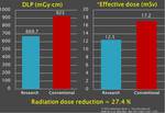
The average DLP for the research protocol was 669.70 mGy‧cm and 923.01 mGy‧cm for the conventional protocol,
equating to average effective doses of 12.46 mSv and 17.17 mSv respectively,
when using E/DLP conversion factor of 18.6 μSv/mGy cm(5) (Table 1).
This corresponds to a radiation dose reduction of 27.44%.
The image quality scores are provided in table 2.
There was poor Interobserver agreement between the two radiologists in regards to the individual scores,
with low kappa values.
However,
when looking at the combined mean score...
Conclusion
There was a significant (27%) patient radiation dose reduction for CT CAP in oncology patients by using a single-pass CT CAP protocol.
The image quality of the single-pass protocol appears to be comparable to the conventional protocol.
The single-pass protocol scans were also not more difficult to perform by radiographers than the conventional protocol scans.
However,
as statedbelow there were significant limitations encountered and more research in this area is required for validation of the results.
Limitations:
Although the radiologists were not informed what protocol...
Personal information
Ernest Lekgabe,
MBBS
Department ofRadiology,
The Alfred,
Melbourne,
Australia.
Ngon Tran
Head CT radiographer,
Department ofRadiology,
The Alfred,
Melbourne,
Australia.
Wa Cheung,
MBBS,
RANZCR
Head of Body Imaging,
Department ofRadiology,
The Alfred,
Melbourne,
Australia.
Ben McDonald,
MBBS,
RANZCR
Department ofRadiology,
The Alfred,
Melbourne,
Australia.
Helen Kavnoudias,
Ph.D.
Department of Radiology,
The Alfred,
Melbourne,
Australia.
Lauren Hudson
Department ofRadiology,
The Alfred,
Melbourne,
Australia.
Xiao Chen,
MBBS
Department ofRadiology,
The Alfred,
Melbourne,
Australia.
References
1.
Yaniv G,
Portnoy O,
Simon D,
Bader S,
Konen E,
Guranda L.
Revised protocol for whole-body CT for multi-trauma patients applying triphasic injection followed by a single-pass scan on a 64-MDCT.
Clin Radiol.
Jul 2013;68(7):668-675.
2.
Nguyen D,
Platon A,
Shanmuganathan K,
Mirvis SE,
Becker CD,
Poletti PA.
Evaluation of a single-pass continuous whole-body 16-MDCT protocol for patients with polytrauma.
AJR Am J Roentgenol.
Jan 2009;192(1):3-10.
3.
Fanucci E,
Fiaschetti V,
Rotili A,
Floris R,
Simonetti G.
Whole body 16-row multislice CT in emergency...


