Learning objectives
The purpose of thiseducational exhibit is to:
provide relevant data on the epidemiology,
pathomorphology and clinical manifestations of this type of CNS tumors;
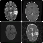
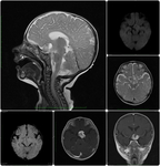
describe and illustrate the MR-imaging findings ofbrain astrocytoma in young children;
describethe use of advanced high-field MRI techniques in the differentiation ofbrain astrocytoma with other conditions;
present our experience in studyingsuch patients in past 3years.
Background
Epidemiology
Astrocytic tumors are the most common brain tumors of childhood,
comprising approximately20% of all childhood malignancies and morethan half of all primary CNS malignancies.
Most cases occur in the first decade of life,
with the peak incidence occurring in children aged 5-9 years.
High-grade supratentorial tumors occur slightly later,
with a median age at diagnosis of 9-10 years.
The term "astrocytoma" containsnumeroustumors that differ by location,
morphologicalcharacteristics,
the degree of invasiveness,
growth rate,
tendency to progression and clinical course.
Pathomorphology
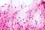
Astrocytomas are the most...
Findings and procedure details
Eight children (mean age 2.1 years,
range 0.2-4.1 years) underwent multiparametric preoperative contrast enhanced MRI in our departmentbetween November2012and August 2015.
All MRIexaminations were performed with a 1.5T MR imaging system (Siemens Magnetom Essenza 1.5T; Siemens Healthcare,
Germany) with a standard head coil.
Intravenous contrast enhancement was made in every case.
In six of eight cases diagnoses of astrocytoma were histologicaly confirmed (two of them were recognized inoperable).
All studies were performed under general anesthesia.
The average total time for research - 28 minutes.
Preoperative...
Conclusion
MRI is the modality of choice for pre-operative and follow-up imaging of brain astrocytomas;
MRI protocol must necessarily include DWI with ADC measurement,
contrast - enhanced3D T1-GREsequensesfor a more accurate differential diagnosis andsuggesting a grade-level with a high degree of probability;
Priority should be given to high-field MRI equipment;
In young children,
the study must be carried out under general anesthesia.
Personal information
Alexey Kokunin M.D.,
radiologist
Regional Diagnostic Center,
MRI Department (Nizhniy Novgorod,
Russia);
Balakhna Central Regional Hospital,
Radiology department(Balakhna,
Russia);
E-mail to contact:
[email protected]
https://linkedin.com/in/alexey-kokunin-500350101
References
MuroffLR,
RungeVM.The use of MR contrast in neoplastic disease of the brain.Top Magn Reson Imaging1995;7:137–57
Louis DN,
Ohgaki H,
Wiestler OD et-al.
The 2007 WHO classification of tumours of the central nervous system.
Acta Neuropathol.
2007;114 (2): 97-109.Acta Neuropathol.
(full text)-doi:10.1007/s00401-007-0243-4-Free text at pubmed-Pubmed citation
Server A,
Kulle B,
Mæhlen J,
et al.
Quantitative apparent diffusion coefficients in the characterization of brain tumors and associated peritumoral edema.Acta Radiol2009; 50:682–689[CrossRef][Medline]
Cha S.Update on brain tumor imaging: from anatomy to physiology.AJNR Am J Neuroradiol2006;27:475–87FREEFull Text
Higano S,...


