Learning objectives
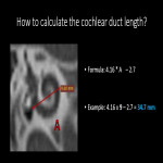
Show anatomy of the internal ear and essential measures for cochlear implants.
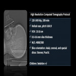
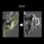
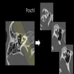
Show appropriate protocol for cochlear implant using a high-resolution computed tomography scan (ultra-low-dose of radiation technique).
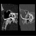
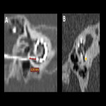
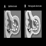
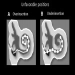
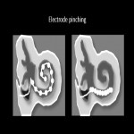
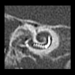
Show the pearl's findings post-implantation of a cochlear implant.
Background
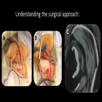
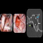
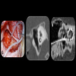
Ear imaging is mainly performed by CT and MRI, each modality has its strengths and limitations. CT is the preferred modality for delineating the inner ear's intricate bony anatomy and malformations. It is important to recognize the key findings in preoperative scans that can impact the surgical procedure. In addition, an understanding of the surgical approach is essential if a postoperative evaluation is to be performed. These educational exhibits explain the essential points for an adequate assessment using computed tomography with low radiation doses and...
Findings and procedure details
Relevant anatomy:
External Auditory canal.
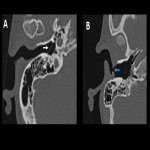
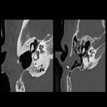
Extends from the auricle to the tympanic membrane, the lateral portion fibrocartilaginous and the medial portion bony.
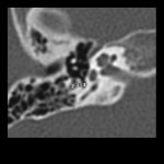
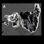
The tympanic membrane normally should only be faintly discernible on CT. A perforation appears as a focal defect. [Fig 1]
Mastoid.
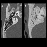
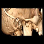
The mastoid air cells are divided by bony septations.
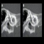
In the central superior aspect is a larger cavity devoid of septations, termed the mastoid antrum. The mastoid antrum communicates with the epitympanum via the aditus ad antrum. [Fig 2]
Middle ear.
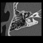
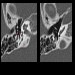
A...
Conclusion
The evaluation of the ear by CT offers an excellent representation of the anatomy of the bony structures of the temporal bone. An adequate acquisition and reconstruction of the images are necessary for a correct interpretation and to be able to guide the otorhinolaryngologist to choose the best approach and the identification of pathologies that have an impact on the surgical procedure. Post-operative imaging is important to document the electrode position and demonstrate findings that may result in poor cochlear implant function.
Personal information and conflict of interest
B. C. Zaragoza:
Nothing to disclose
O. D. GARCIA:
Nothing to disclose
References
Amy F. Juliano, ‘Cross-Sectional Imaging of the Ear and Temporal Bone, Head and Neck Pathology, 12.3 (2018), 302 <https://doi.org/10.1007/S12105-018-0901-Y>.
Joshi VM and others, ‘CT and MR Imaging of the Inner Ear and Brain in Children with Congenital Sensorineural Hearing Loss’, Radiographics : A Review Publication of the Radiological Society of North America, Inc, 32.3 (2012), 683–98 <https://doi.org/10.1148/RG.323115073>.
Gerlig Widmann and others, ‘Pre- and Post-Operative Imaging of Cochlear Implants: A Pictorial Review’, Insights into Imaging (2020) <https://doi.org/10.1186/s13244-020-00902-6>.
Thomas Lenarz, ‘Cochlear Implant – State of the Art’,...


