Aims and objectives
Although HIV replication in the central nervous system (CNS) is generally controlled by systemic combined antiretroviral therapy (cART),
cases of encephalitis associated with cerebrospinal fluid (CSF) viral escape and resembling old HIV-encephalitis (HIV-E) cases of untreated patients are increasingly being observed.Cerebrospinal Fluid (CSF) viral escape,
i.e.,
detectable HIV-RNA in CSF despite undetectable or lower-than-CSF level in plasma,
is seen in approximately 10% of patients receiving combination antiretroviral treatement (cART)[1].
This condition is infrequent,
although individual case descriptions and small case series are increasingly being reported...
Methods and materials
We retrospectively analyzed radiological,
clinical and virological data of 21HIV-infected patients with CSF escape encephalitis,
i.e.,
incident neurological symptoms while on ART (>9 months) and detectable CSF HIV-RNA with concomitant,
undetectable plasma HIV-RNA,
or CSF HIV-RNA higher than in plasma,
and in 17 patients with classical HIV-E.Although all patients were analyzed retrospectively and not all have done MRI in our center we considered for the morphologic evaluation presence of signal alterationin T1,
T2 FLAIR and DWI sequences.
For these reasons MR images were totally disposable...
Results
Patients characteristics and laboratory findings at the time of diagnosis of CSF escape or HIV-encephalitis are reported in Table 1.
Patients clinical symptoms and signs are graphically reported in Table 2.
Neurological presentation consisted of memory and cognitive impairment (HIV-E=10; CSF escape=10),
agitation/psychosis/delusions (HIV-E=4; CSF escape=0),
alteration of consciousness (HIV-E=3; CSF escape=4),
cerebellar signs (HIV-E=3; CSF escape=11),
focal deficits (HIV-E=2; CSF escape=8).
MRI findings are graphicallyreported in Table 3.
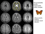
In classical HIV-E,
MRIshowed diffuse bilateral periventricular white matter hyperintensity (Fig 1),
most frequently symmetrical involving...
Conclusion
Despite similarities in clinical presentations,
specific MRI and CSF findings were observed in esc-HIVE compared to pre-cART HIVE in particular the presence of frequent inflammatory pattern at MRI,
with no atrophy.
Primarily diffuse periventricular white matter hyperintensity,
CSF escape differed from classical HIV-E for a more frequent involvement of corpus callosum,
cerebellar regions and brainstem and,
more in general,
for the presence of reversible edema with mass effect.
From a viral point of view lower CSF levels of HIV replication and higher CSF cell numbers...
Personal information
Simonetta Gerevini,
M.D.
Neuroradiological Unit,
IRCCS San Raffaele Hospital,
MI,
Italy.
Phone +390226433028
Fax +390226433447
email:
[email protected]
Paola Cinque,
M.D.
PhD.
Department of Infectious Diseases IRCCS San Raffaele Hospital,
MI,
Italy.
Phone +390226433160
email:
[email protected]
References
1.
Ferretti F,
Gisslen M,
Cinque P,
Price RW.Cerebrospinal Fluid HIV Escape from Antiretroviral Therapy.Curr HIV/AIDS Rep2015; 12:280-8.
2.
Canestri A,
Lescure F-X,
Jaureguiberry S,
Moulignier A,
Amiel C,
Marcelin AG,et al.Discordance between cerebral spinal fluid and plasma HIV replication in patients with neurological symptoms who are receiving suppressive antiretroviral therapy.Clin Infect Dis2010;50:773-8.
3.
Peluso MJ,
Ferretti F,
Peterson J,et al.Cerebrospinal fluid HIV escape associated with progressive neurologic dysfunction in patients on antiretroviral therapy with well controlled plasma viral load.AIDS2012;26:1765–1774
4.
Kugathasan R,
Collier DA,...

; in some case edema with sulcal effacement can be seen depending of the degree of the underlying atrophy (lower line)")

